DermNet provides Google Translate, a free machine translation service. Note that this may not provide an exact translation in all languages

Translate

Subungual melanoma pathology

Author: Adjunct A/Prof Patrick Emanuel, Dermatopathologist, Clínica Ricardo Palma, Lima, Peru. DermNet NZ Editor in Chief: Adjunct A/Prof Amanda Oakley. Copy edited by Maria McGivern/Gus Mitchell. June 2018. 


toc-icon
Table of contents
arrow-right-small

Introduction

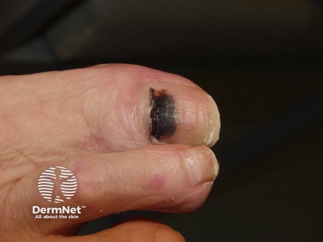
Subungual melanoma is a cancer that arises from a malignant proliferation of melanocytes in the nail matrix. Typically, it presents clinically as a pigmented streak in the nail plate, which slowly expands at the proximal border and may extend to involve the adjacent nail fold (Hutchinson sign; figure 1).

Subungual melanoma

Histology of subungual melanoma

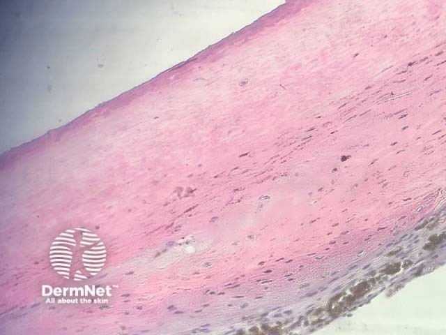
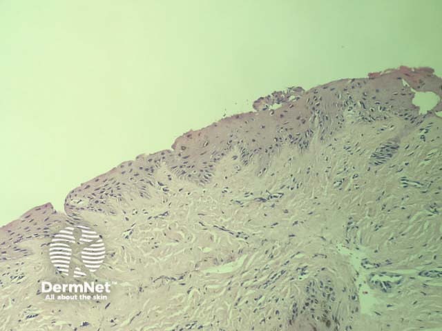
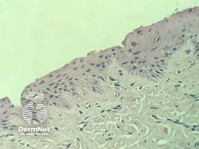
Histopathologically, the nail plate in subungual melanoma will show abundant diffuse melanin deposition (figure 2). Sections of the matrix and nail bed show a melanocytic lesion with poor circumscription, predominantly single-cell growth, and pagetoid scatter (figures 3 and 4). Junctional nesting and confluence may be seen. The majority of cases demonstrate a combination of epithelioid and dendritic cytomorphology (figure 5). Nuclear atypia and conspicuous nucleoli are often overt but may be subtle.  

Subungual melanoma

Special studies for subungual melanoma

Immunohistochemistry using Melan-A and SOX10 can be useful in highlighting the extent and pattern of the melanocytic lesion and help to confirm or rule out invasion. Proliferative markers (eg, Ki-67) can be used to highlight an increased proliferative index.

Fluorescent in-situ hybridisation (FISH) may be used to differentiate melanoma from benign melanocytic lesions

Differential diagnosis for subungual melanoma

Other diagnoses to be considered include:

  • Subungual atypical lentiginous melanocytic proliferation — this moniker has been proposed for difficult cases that don’t fulfil all the criteria for melanoma, particularly in paediatric cases; it is generally recommended that these borderline cases be treated as melanoma (FISH may be used to help illustrate an aneuploid clone, which is likely to be malignant).
  • Matrical naevi and lentigines — these lack the atypical histopathological features seen in melanoma. 


References

  1. Khatri SS, Wang M, Harms KL, et al. Subungual atypical lentiginous melanocytic proliferations in children and adolescents: a clinicopathologic study. J Am Acad Dermatol 2018; 79: 327–36.e2. DOI: 10.1016/j.jaad.2018.03.030. PubMed
  2. Kleinerman R, Kriegel D, Amir I, Emanuel PO, Markinson BC. Osteoinvasive subungual melanoma: a case and review. J Drugs Dermatol 2010; 9: 159–63. PubMed

On DermNet

Other websites

Books about skin diseases

Text: Miiskin


Related information

Sign up to the newsletter