

Systemic amyloidosis pathology — extra information
Introduction
Histology
Special studies
Differential diagnoses
Introduction
Systemic amyloidosis caused by the buildup of amyloid in various organs. More than 30 different proteins can cause systemic amyloidosis and the causes are diverse.
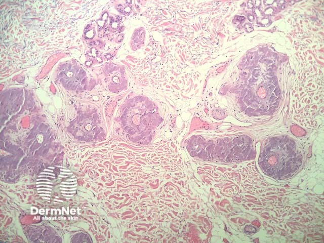
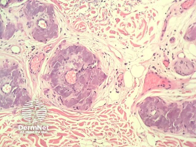
Histology of systemic amyloidosis
In systemic amyloidosis, the histopathology shows pale pink, extracellular, glassy, hyaline material mostly deposited in and around vessels (figures 1,2).
Special studies for systemic amyloidosis
Amyloid stains positively with congo red, periodic acid–Schiff (PAS), crystal violet, and toluidine blue. Characteristically, the congo red stain produces an apple-green birefringence under polarised microscopy.
Differential diagnosis for systemic amyloidosis
Other conditions that should be considered include:
- A form of primary cutaneous amyloidosis — the most important differential. Keratin immunohistochemistry is usually positive in lichen amyloidosis and macular amyloidosis. A primary vascular location of deposition points to systemic amyloidosis
- Erythropoietic protoporphyria — hyaline deposits are seen around and within superficial blood vessel walls. Involvement of the dermoepidermal junction may be seen
- Lipoid proteinosis — dermal deposits are also accentuated around blood vessels, however the material is usually weakly positive or negative for congo red. Clinical correlation can be very helpful.
References
- Pathology of the Skin (Fourth edition, 2012). McKee PH, J. Calonje JE, Granter SR.
On DermNet
- Systemic amyloidosis
- Cutaneous amyloidosis
- Macular amyloidosis
- Dermatopathology glossary
- Dermatopathology index