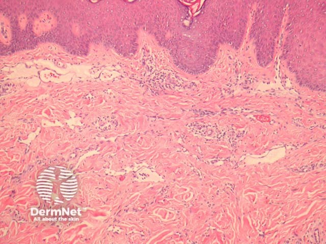
Also known as hobnail haemangioma and targetoid haemangioma, targetoid haemosiderotic haemangiomas are rare angiomas and present clinically as erythematous or violaceous papules or macules surrounded by a distinctive pale or dark ring (imparting a targetoid appearance).
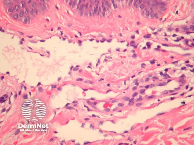
In targetoid haemosiderotic haemangioma, the surface of the lesion shows ectatic vessels lined by prominent epithelioid endothelial cells (“hobnail” endothelial cells). See figures 1,2.
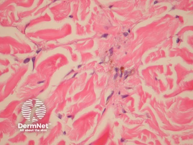
Deeper in the dermis, the endothelial cells are less prominent and form slit-like spaces through the collagen fibres. Haemosiderin deposition is common. See figure 3.
The vascular nature of the lesion can be confirmed with immunohistochemical studies with CD31 or CD34.
Angiosarcoma: The prominent endothelial cells can be confused with angiosarcoma. Lack of significant nuclear atypia, minimal infiltration of deeper tissues, and low proliferative index favour targetoid haemosiderotic haemangioma.
Kaposi sarcoma: Deep aspects of targetoid haemosiderotic haemangioma form slit-like spaces between collagen fibres which resemble Kaposi sarcoma. Kaposi sarcoma generally does not show “hobnail” endothelial cells. Immunohistochemistry for HHV-8 can be helpful in confirming suspected cases of Kaposi sarcoma.

