DermNet provides Google Translate, a free machine translation service. Note that this may not provide an exact translation in all languages

Translate

Transient neonatal pustular melanosis

Author: Dr Diana Norris, Dermatology Registrar, Skin and Cancer Foundation Victoria, Carlton, VIC, Australia. DermNet New Zealand Editor in Chief: Adjunct A/Prof Amanda Oakley, Dermatologist, Hamilton, New Zealand. Copy edited by Gus Mitchell/Maria McGivern. February 2018. 


toc-icon

What is transient neonatal pustular melanosis?

Transient neonatal pustular melanosis is an uncommon benign pustular condition presenting in newborn infants [1]. It is also known as transient neonatal pustular dermatosis and transient neonatal pustulosis.

Neonatal pustular melanosis

Who gets transient neonatal pustular melanosis?

Transient neonatal pustular melanosis affects 0.2–4% of newborn infants in the first few days of life [2]. It is more common in African American babies than white American babies, affecting 4.4% of African American [3].

What causes transient neonatal pustular melanosis?

The cause of transient neonatal pustular melanosis is unknown. Some authors have suggested it may be a variant of toxic erythema of the newborn [4].

What are the clinical features of transient neonatal pustular melanosis?

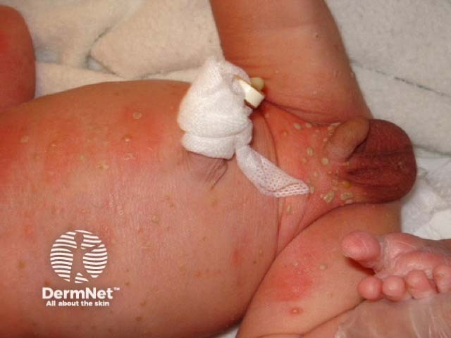
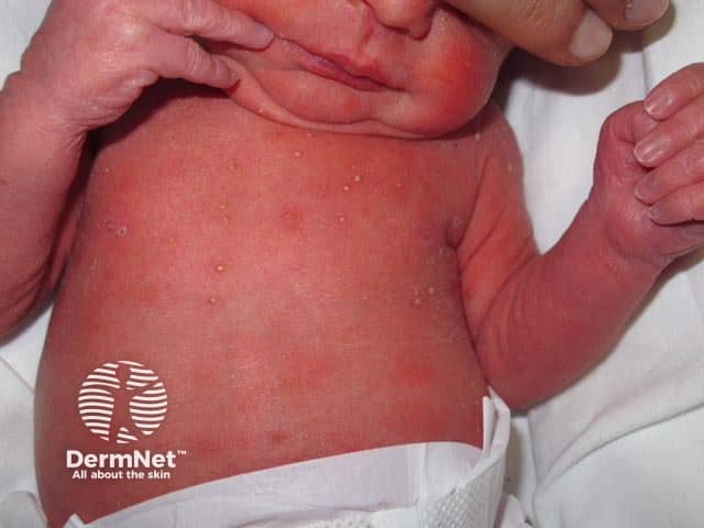
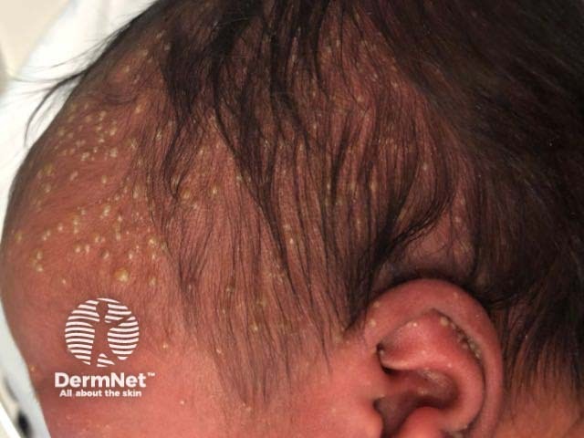
The key clinical feature of transient neonatal pustular melanosis is the presence of pustules. These occur on an unaffected, non-erythematous base [1].

  • Any area can be involved, including the patient's forehead, posterior ears, chin, neck, upper chest, back, buttocks, abdomen, thighs, palms, and soles [1].
  • Lesions can be either solitary or occur in clusters [1].
  • The pustules can rupture and crust, resolving within several days [1].
  • The pustules may leave an area of hyperpigmentation, which fades within weeks to months [1].
  • The pigmented macules can have a collarette of scale [2].
  • The pustules are present at birth and evolve; new lesions usually do not develop after birth [1].
  • There are no associated systemic symptoms or laboratory abnormalities.

How is transient neonatal pustular melanosis diagnosed?

The diagnosis of transient neonatal pustular melanosis is clinical. No investigations are required.

skin biopsy shows a neutrophilic infiltrate in the epidermis or dermis, commonly with subcorneal pustules, and occasional eosinophils [2,5]. Pustules contain neutrophils with no organisms [1].

What is the differential diagnosis for transient neonatal pustular melanosis?

The differential diagnosis for transient neonatal pustular melanosis includes:

What is the treatment for transient neonatal pustular melanosis?

No treatment is necessary for transient neonatal pustular melanosis, as it is self-resolving and has no long-term complications [5,6].


References

  1. Schachner LA, Hansen RC, B Krafchik, et al (eds). Pediatric dermatology, 4th ed. Maryland Heights: Mosby, 2011.
  2. Merlob P, Metzker A, Reisner SH. Transient neonatal pustular melanosis. Am J Dis Child 1982; 136: 521–2. PubMed.
  3. Brazzelli V, Grasso V, Croci G, Figar T, Borroni G. An unusual case of transient neonatal pustular melanosis: a diagnostic puzzle. Eur J Pediatr 2014; 173: 1655–8. DOI: 10.1007/s00431-013-2227-9. PubMed.
  4. Ferrándiz

    C, Coroleu LW, Ribera M, Lorenzo JC, Natal A. Sterile transient neonatal pustulosis is a precocious form of erythema toxicum neonatorum. Dermatology 1992; 185: 18–22. DOI: 10.1159/000247396. PubMed.
  5. Elder D, Elenitsas R, Jaworsky C, Johnson B Jr (eds). Lever’s histopathology of the skin, 8th edn. Philadelphia: Lippincott-Raven, 1997.
  6. Van Praag M, Van Rooij R, Folkers E, Spritzer R, Menke H, Oranje A. Diagnosis and treatment of pustular disorder in the neonate. Pediatr Dermatol 1997; 14(2): 131–43. PubMed.

On DermNet

Other websites

Books about skin diseases


Related information

Sign up to the newsletter