DermNet provides Google Translate, a free machine translation service. Note that this may not provide an exact translation in all languages

Translate

Trichilemmal cyst

Author: Anila Kapadia, Dermatology Registrar, York Teaching Hospital NHS Foundation Trust, United Kingdom. Advisor to the content: Dr. Calum Lyon, Consultant Dermatologist, York Teaching Hospital NHS Foundation Trust, United Kingdom. Copy Editor: Clare Morrison. June 2014.


toc-icon

What is a trichilemmal cyst?

A trichilemmal cyst, also known as a pilar cyst, is a keratin-filled cyst that originates from the outer hair root sheath. Keratin is the protein that makes up hair and nailsTrichilemmal cysts are most commonly found on the scalp and are usually diagnosed in middle-aged females. They often run in the family, as they have an autosomal dominant pattern of inheritance (ie, the tendency to the cysts can be is passed on by a parent to their child of either sex, and the child has a 1 in 2 likelihood of inheriting it).

What are the clinical features of trichilemmal cyst?

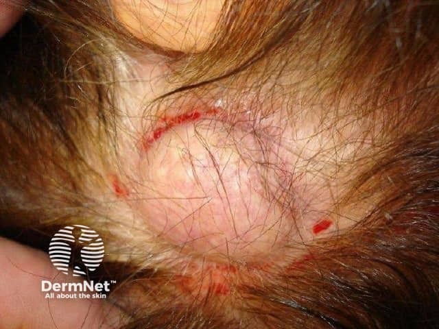
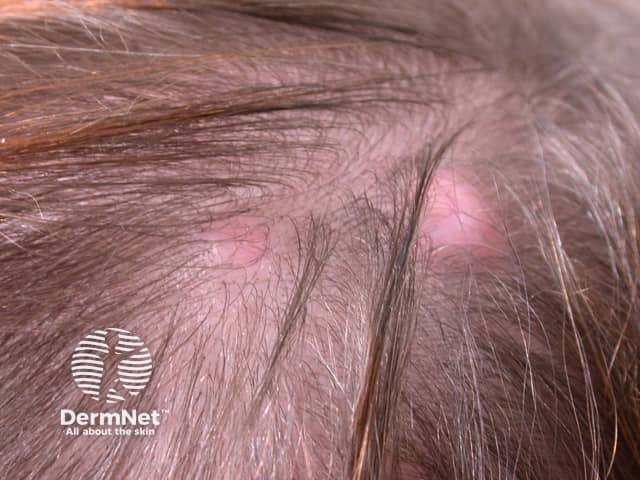
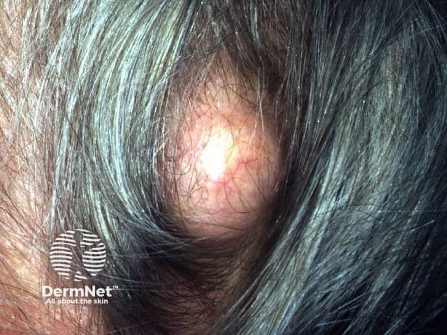
Trichilemmal cysts may look similar to epidermoid cysts and are often incorrectly termed sebaceous cysts. Trichilemmal cysts present as one or more firm, mobile, subcutaneous nodules measuring 0.5 to 5 cm in diameter. There is no central punctum, unlike an epidermoid cyst. A trichilemmal cyst can be painful if inflamed.

Pilar cyst

More images of cysts ...

How does trichilemmal cyst differ from epidermoid cyst?

Trichilemmal cyst

  • Most common sites for trichilemmal cyst are scalp (90%) and scrotum.
  • Central punctum is absent.
  • Origin is outer root sheath.
  • Cyst wall is thick and not prone to rupture.
  • Histology: granular layer is absent.

Epidermoid cyst

  • Most common sites for epidermoid cyst are face, neck and trunk.
  • Central punctum is present.
  • Origin is epithelium or hair follicle infundibulum.
  • Cyst wall is delicate and prone to rupture.
  • Histology: granular layer is present  

What are the histological findings in trichilemmal cysts?

The pathology of a trichilemmal cyst is characteristic. The wall of the cyst is stratified squamous epithelium (skin) that has a palisaded outer layer, which resembles the that of outer root sheath of a hair follicle. The inner layer does not have a granular layer. The cyst shows very dense pink keratin on haematoxylin and eosin staining.

What is the treatment for trichilemmal cysts?

It is not necessary to remove trichilemmal cysts if they are not causing symptoms. However, incision and drainage under local anaesthesia provide comfort, and elective excision before rupture prevents scarring.

Surgical treatment involves either of the following methods:

  • Enucleation of the cyst, ie removal without cutting into it and leaving surrounding skin intact
  • Incision followed by expression of contents and removal of cyst wall – this is often best achieved with a surgical punch with the cyst expressed through the hole.

Acute inflammation after rupture is often misdiagnosed as a bacterial infection. Antibiotics are of little value unless an actual infection is present.


References

  • Eds Burns T, Breathnach S, Cox N, Griffiths C. Rook’s Textbook of Dermatology. 8th edition 2010. Wiley-Blackwell Publications.
  • Velez AMA, Brown VN and Howard MS. An inflamed trichilemmal (pilar) cyst: Not so simple? North Am J Med Sci 2011; 3: 431–4. PubMed

On DermNet

Books about skin diseases


Related information

Sign up to the newsletter