Introduction Demographics Causes Clinical features Complications Diagnosis Differential diagnoses Treatment Outcome
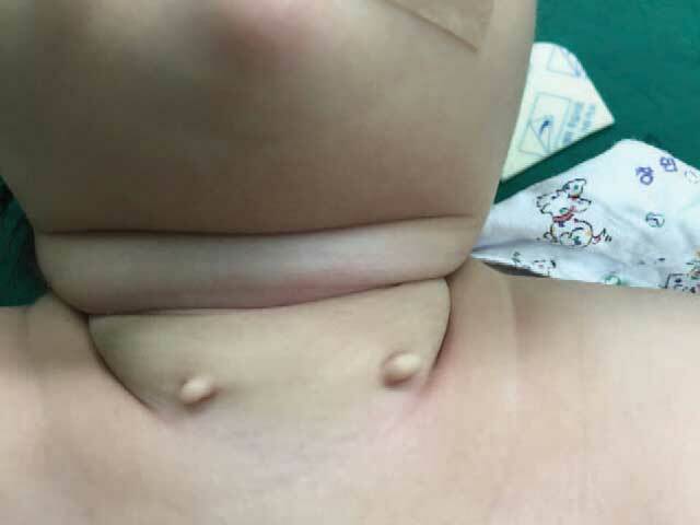
Congenital cartilaginous rests of the neck are rare, benign, congenital anomalies that likely represent branchiogenic malformations. They often present unilaterally, and less commonly, bilaterally over the anterior neck.
These appendages were first termed ‘wattles’ due to their similarity in appearance to dewlaps in birds, such as those seen in turkeys and chickens. Many domestic goats, pigs, and sheep also have wattles.
Congenital cartilaginous rests of the neck are also referred to as cervical chondrocutaneous branchial remnants.
Congenital cartilaginous rests of the neck are believed to originate from the first or second branchial arch or auricular tissues.
During the fourth week of embryogenesis, neural crest cells migrate to the future head and neck, forming six branchial arches. Each arch comprises an internal endodermal layer, mesenchymal core, and outer ectodermal layer. By the seventh week of embryogenesis, the auricles have developed from the auricular hillocks, derived from the first and second branchial arches. On route to their final position on the head, the developing auricles may leave cartilaginous deposits along their migratory path which overlies the sternocleidomastoid muscle. If this occurs, there will be a permanent structural anomaly over the anterolateral neck in the case of congenital cartilaginous rests of the neck or anterior to the auricle in the case of an accessory auricle (accessory tragus or preauricular appendage).
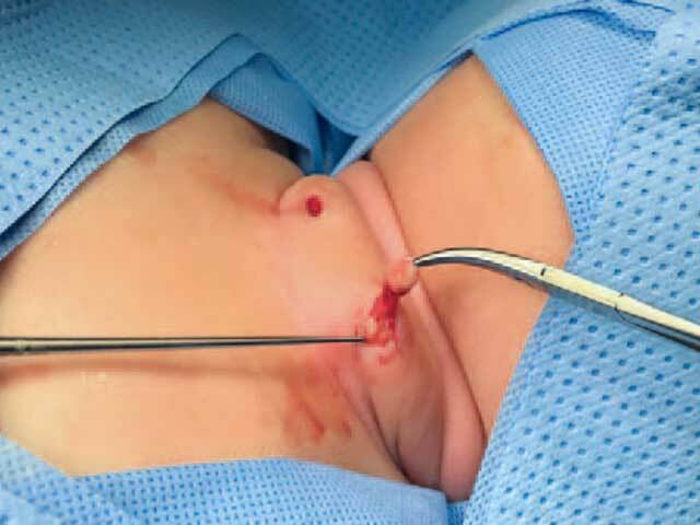
Congenital cartilaginous rests of the neck typically present as a solitary sessile or pedunculated, skin-coloured papule or nodule. Lesions are firm to palpation and measure between 5 to 25 mm in size. They are usually located over the middle or lower third of the lateral neck anterior to the sternocleidomastoid muscle. The overlying skin is unremarkable and not inflamed, and lesions are typically asymptomatic, demonstrating no or very slow growth.
Congenital cartilaginous rests of the neck often adhere to the fascia of the sternocleidomastoid muscle, although they do not connect with deeper structures. There are no reports of underlying sinuses or cysts.
Congenital cartilaginous rests of the neck are benign lesions; however, they may be associated with underlying cardiac, genitourinary, auditory, respiratory, gastrointestinal, musculoskeletal, and ocular anomalies.
Published case series report one or more underlying congenital anomalies in 11% to 76% of cases with well-characterised congenital syndromes including Treacher-Collins, Goldenhar, and Nager also associated with the condition. It is therefore recommended that a complete systems enquiry, thorough examination, and ultrasonographic imaging of the heart and abdomen be undertaken.
The diagnosis of congenital cartilaginous rests of the neck can usually be made clinically. However, ultrasonography can help to confirm the clinical suspicion before surgical excision. The ultrasonographic presence of tubular cartilage extending to the sternocleidomastoid muscle is characteristic.
A complete excision/skin biopsy would provide a definitive histologic diagnosis demonstrating tubular hyaline or elastic cartilage covered by normal skin consisting of an intact and unremarkable epidermis, dermis, and subcutaneous adipose layer.
For cosmetic reasons and to obtain an accurate histologic diagnosis, congenital cartilaginous rests of the neck are best treated by complete surgical excision.
Congenital cartilaginous rests of the neck are rare benign lesions that do not recur following surgical excision. The presence of co-existing congenital anomalies should be considered in all patients diagnosed with this condition.

