

Cutaneous B-cell lymphoma — extra information
Introduction - lymphoma
Introduction - cutaneous B-cell lymphoma
Demographics
Skin signs
Diagnosis
Treatment
Outcome
What is lymphoma?
Lymphomas are tumours of the lymph nodes and lymphatic system.
- Extranodal lymphomas are tumours that occur in organs or tissues outside of the lymphatic system.
- Primary cutaneous lymphomas occur in the skin with no evidence of extracutaneous disease at the time of diagnosis.
Primary cutaneous lymphoma can be broadly divided into two categories:
- Cutaneous T-cell lymphoma
- B-cell lymphoma.
Primary cutaneous B-cell lymphomas (PCBCL) comprise approximately 20% of cutaneous lymphomas and are described on this page.
What are cutaneous B-cell lymphomas?
Cutaneous B-cell lymphomas are a malignant proliferation of lymphocytes of the B-cell type. Mutation occurring at different points in B cell development leads to different forms of lymphoma.
In 2005, the World Health Organization (WHO) and the European Organization for Research and Treatment of Cancer Classification (EORTC) reached a consensus classification for cutaneous lymphomas. It was revised by the WHO in 2008. The three main types of PCBCL are:
- Primary cutaneous follicle centre lymphoma (PCFCL)
- Primary cutaneous marginal zone B-cell lymphoma (PCMZL)
- Primary cutaneous diffuse large B-cell lymphoma, leg type (PCDLBCL-LT).
Rare cases of PCDLBCL that do not belong to the PCDLBCL-LT or PCFCL groups are categorised as PCDLBCL-other. Other entities such as anaplastic or plasmablastic lymphoma, primary cutaneous T-cell/histiocyte-rich B-cell lymphomas and primary cutaneous intravascular large B-cell lymphomas are very rare.
Due to differences in treatment and prognosis, it is important to discern between PCBCL and systemic forms of B-cell lymphomas that manifest with secondary skin involvement.


Who gets primary cutaneous B-cell lymphoma?
Primary cutaneous follicle centre lymphoma
- Primary cutaneous follicle centre lymphoma (PCFCL) is the second most common lymphoma in the Western world.
- It is the most common type of PCBCL.
Primary cutaneous marginal zone B-cell lymphoma
See primary cutaneous marginal zone B-cell lymphoma (PCMZL).
- Men are affected twice as often as women.
- Some are associated with Borrelia burgdorferi infection (the cause of Lyme disease) in endemic areas of Europe and the USA.
Primary cutaneous diffuse large B-cell lymphoma, leg type (PCDLBCL-LT)
- PCDLBCL-LTT is twice as common in women than in men.
- It has the worst prognosis, with 5-year survival 50%.
- It affects older people with median age 76 years.
Primary cutaneous diffuse large B-cell lymphoma, other (PCDLBCL-other)
- PCDLBCL-other is very rare.
- It has a variable prognosis.
What are skin signs of primary cutaneous B-cell lymphoma?
Primary cutaneous follicle centre lymphoma
- PCFCL presents with solitary or grouped papules, plaques, or nodules.
- These are pink to violaceous.
- They are usually found on scalp, forehead, or trunk.
- They are very rarely found on the leg.
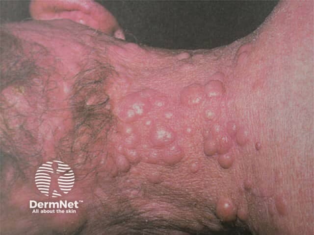
Primary cutaneous marginal zone B-cell lymphoma
PCMZL is a low-grade malignant B-cell lymphoma of the MALT (mucosa-associated lymphoid tissue) type.
- PCMZL presents with solitary or multiple papules, plaques or nodules.
- They are red to violaceous.
- They are most often found on the trunk and upper extremities.
Primary cutaneous diffuse large B-cell lymphoma, leg type
- PCDLBCL-LTT presents with solitary or multiple nodules, infiltrated plaques and tumours.
- Rarely, it presents as verrucous plaques or widespread garland-like lesions.
- They are typically red to bluish.
- They are mainly found on one or both legs but it affects other sites in 10–15%.
- It represents an aggressive subtype of PCBCL.
- Multiple skin lesions on the leg have a poor prognosis.
Primary cutaneous diffuse large B-cell lymphoma, other
- PCDLBCL-other includes large B-cell lymphoma, which presents with skin lesions on the head, the trunk or the extremities.
- Blastic plasmacytoid dendritic cell neoplasm presents with bruise-like plaques, nodules and tumours. It later progresses to involve the central nervous system. It represents a type of leukaemia cutis.
- Intravascular large B-cell lymphoma proliferates within the lumen of small blood vessels, primarily in the skin and central nervous system and later other organs. Lesions appear as erythematous, tender nodules, tumours, and telangiectases mainly on the trunk and lower legs. Cutaneous lesions may be confused with mycosis fungoides, sarcoidosis, blood vessel tumours, or leukaemia cutis.


How is cutaneous B-cell lymphoma diagnosed?
The work-up of PCBCL should include a complete history, physical and skin examination. Initial blood tests should include:
- Full blood count and differential
- Metabolic profile and lactate dehydrogenase.
If the full blood count shows lymphocytosis, peripheral blood flow cytometry may be needed.
An adequate skin biopsy is also important for evaluation and staging. Either 4–6 mm punch, incisional, or excisional biopsies should be obtained and should include the reticular dermis and subcutaneous fat. Superficial biopsy specimens may not differentiate PCBCL from reactive or inflammatory processes.
Assessment for extra-cutaneous involvement should be done, particularly in patients with palpable lymphadenopathy.
- Contrast CT scan
- PET scan
Any lymph node identified on imaging > 1.5 cm in length or showing high PET activity should undergo a biopsy; an excisional biopsy is preferred where possible.
Histologically, PCBCL shows a diffuse monotonous population of centroblasts and immunoblasts. Histochemistry tests are essential to classify the exact type of lymphoma.
Primary cutaneous follicle centre lymphoma
PCFCL needs to be differentiated from a secondary cutaneous lymphoma, which is where a nodal follicular lymphoma has spread to involve the skin. Cellular morphology may vary with the age and size of the lesion.
Histologically, PCFCL shows:
- Dermal and subcutaneous proliferation of centrocytes (cleaved follicle centre cells) and centroblasts (large transformed cells) in a follicular and/or diffuse growth pattern.
Immunostaining shows:
- Positive B-cell antigens, ie CD20 and CD79a
- Negative bcl-2 protein, and, the t(14;18) translocation (which if present may suggest systemic follicular lymphoma)
- Negative immunoglobulin M expression.
A FISH analysis is unhelpful.
Primary cutaneous marginal zone B-cell lymphoma
The histology of PCMZL consists of:
- Nodular to diffuse dermal infiltrates of marginal zone B-cells, lymphocytes, lymphoplasmacytoid cells, plasma cells admixed with reactive T cells and centroblast or immuneblast-like cells
- Marginal zone B-cells, which tend to be small to medium in size with irregular nuclei, inconspicuous nucleoli and abundant pale cytoplasm. They proliferate in the dermal and subcutaneous regions of skin.
Immunostaining shows
- Positive CD20, CD79a and Bcl-2
- Negative CD10 and Bcl-6.
- Less than 25% of PCMZL harbor the t(14;18) translocation.
- Expresses microRNAs 150 and 155 which, if present, may be predictive of longer progression-free survival.
Primary cutaneous large B-cell lymphoma, leg type
PCFCL and PCDLBCL-LT have diffuse large B-cell infiltrates. PCDLBCL-LT shows:
- Diffuse dermal proliferation of centroblasts and immunoblasts in monotonous or confluent sheets
- Larrge round nuclei with open chromatin, prominent nucleoli and identifiable mitotic figures
- Sparse T cells confined to perivascular areas.
Histopathological differential diagnoses should include:
- Diffuse large B-cell lymphoma not otherwise specified (DLBCL NOS)
- Epstein–Barr virus-positive DLBCL of the elderly
- Lymphomatoid granulomatosis.
Immunostaining shows:
- Positive CD20 and CD79a with strong expression of surface and cytoplasmic immunoglobulin M.
- Strong expression of Bcl-2, MUM1/IRF4 and FOX-P1
- Any PCBCL showing both Bcl-2 and MUM1 expression should be classified as leg type, regardless of anatomic location.
The FISH analysis shows translocations of myc, Bcl-6 and immunoglobulin H genes.
What is the treatment for primary cutaneous B-cell lymphoma?
The rarity of PCBCL and lack of comparative prospective, randomised studies limits the choice of therapy as most treatments are based on data from small retrospective studies. The European Organisation for Research and Treatment of Cancer Cutaneous Lymphoma Group (EORTC-CLG) and the International Society for Cutaneous Lymphoma (ISCL) have uniform recommendations on treating the three main types of PCBCL. Treatment is also influenced by whether the lesion is single or limited to a single site.
Primary cutaneous follicle centre lymphoma
Without any treatment, PCFCL lesions may be stable, gradually enlarge, or rarely, regress. The histological growth pattern does not influence survival or treatment choice.
- If PCFCL presents with a solitary lesion or is limited to a single site, treatment involves radiation therapy or surgery. Skin recurrences are common and often are outside the treated site but these do not affect prognosis.
- Patient with multiple lesions can be observed or given radiation, topical agents, cryotherapy, intralesional steroids or systemic therapy. Where disseminated skin lesions or large tumours occur, chemotherapy may be required such as ie, CHOP or R-CHOP.
- Immune-based therapies like rituximab, interferon and imiquimod are being explored.
Primary cutaneous marginal zone B-cell lymphoma
The recommended therapies for PCMZL are similar to PCFCL.
- Radiotherapy is highly effective and potentially curative for solitary or few contiguous lesions, but the relapse rates of PCMZL can be higher than PCFCL.
- Primary surgical excision can be effective for solitary or localised PCMZL.
- Local treatments include topical corticosteroids, intralesional steroids, topical nitrogen mustard, intralesional rituximab and cryotherapy.
- Multifocal skin lesions are treated with chemotherapy agents such as chlorambucil, interferon-alpha and anti-CD20 antibody (rituximab).
- PCMZL associated with Borrelia burdorferi infection may be treated with antibiotics
- Rarely, disseminated skin lesions are treated with chemotherapy (eg, CHOP).
Primary cutaneous large B-cell lymphoma, leg type
Localised or solitary leg-type B-cell lymphoma is usually are treated with local radiotherapy alone or in conjunction with R-CHOP.
Generalised PCDLBCL-LT can be treated with R-CHOP and/or local radiotherapy. Response rates are high but relapse rates are > 58% Around 30% develop extracutaneous disease.
Trials are assessing the efficacy of biological agents like ofatumumab, lumilixumab, dacetuzumab and intralesional TG1042 in the treatment of PCDLBCL-LT.
What is the outcome for primary cutaneous B-cell lymphoma?
Primary cutaneous follicle centre lymphoma
- Solitary or multifocal PCFCL may enlarge slowly or spontaneously resolve.
- Recurrence occurs in about 46.5% of patients.
- 5-year survival rates are around 95%.
- Dissemination to extracutaneous sites occurs in about 5–10% of cases.
- PCFCL on the leg has a poorer prognosis with 41% survival rate within 5-years.
Primary cutaneous marginal zone B-cell lymphoma
- 5-year disease-specific survival of PCMZL is up to 99%.
- Occasionally, early spontaneous resolution of PCMZL can occur.
- The disease can recur in about 40% of patients, especially if multiple sites are involved.
- It is rare for PCMZL to metastasise to extra-cutaneous sites.
Very rarely, PCMZL can transform into DLBCL.
Primary cutaneous large B-cell lymphoma, leg type
- PCDLBCL-LT frequently disseminates to extracutaneous sites and has a poor prognosis in spite of aggressive therapy.
- 5-year survival rates are approximately 50%.
- In a large multi-centre French study, leg tumours had a 3-year disease-specific survival rate of 43% versus 77% in patients with PCDLBCL not involving the leg.
- 3-year disease-specific survival of patients with multiple skin lesions was 39% compared to 77% in those with single lesions.
Primary cutaneous diffuse large B-cell lymphoma, other
- Large B-cell lymphoma has an excellent prognosis.
- Blastic plasmacytoid dendritic cell neoplasm and intravascular large B-cell lymphoma have a poor prognosis.
References
- Suarez AL, Querfeld C, Horwitz S, Pulitzer M, Moskowitz A, Myskowski PL. Primary cutaneous B-cell lymphomas: Part I Clinical features, diagnosis and classification. J Am Acad Dermatol 2013; 69(3): 329.e1-13. PubMed
- Suarez AL, Querfeld C, Horwitz S, Pulitzer M, Moskowitz A, Myskowski PL. Primary cutaneous B-cell lymphomas: Part II Therapy and Future Directions. J Am Acad Dermatol 2013; 69(3): 343.e1-11. PubMed
On DermNet
- Lymphoma
- Primary cutaneous follicle centre lymphoma
- Primary cutaneous marginal-zone lymphoma
- Primary cutaneous diffuse large B-cell lymphoma — pathology
- Intravascular B cell lymphoma
- Lymphomatoid granulomatosis
- Epstein–Barr virus-associated lymphoproliferative disorders
- Skin lesions, tumours and cancers
- Cutaneous T-cell lymphoma
- Lymphocytoma cutis
- Leukaemia cutis
- Hodgkin lymphoma
- Skin manifestations of haematological disease
Other websites
- Medscape
- Cutaneous Lymphoma Foundation
- Leukaemia & Blood Foundation — New Zealand
- lymphomainfo.net (facts, resources and community)