

Necrotising fasciitis — extra information
Introduction
Causes
Pathophysiology
Clinical features
Diagnosis
Treatment
Outcome
What is necrotising fasciitis?
Necrotising fasciitis is a very serious bacterial infection of the soft tissue and fascia. The bacteria multiply and release toxins and enzymes that result in thrombosis in the blood vessels. The result is the destruction of the soft tissues and fascia.
The main types of necrotising fasciitis are:
- Type I (polymicrobial ie, more than one bacteria involved)
- Type II (due to haemolytic group A streptococcus, and/or staphylococci including methicillin-resistant strains/MRSA)
- Type III (gas gangrene eg, due to clostridium)
- Other: marine organisms (vibrio species, Aeromonas hydrophila, considered Type III in some reports) and fungal infections (candida and zygomycetes, type IV in some reports).
Type I necrotising fasciitis
Bacteria causing type 1 necrotising fasciitis include Staphylococcus aureus, Haemophilus, Vibrio and several other aerobic and anaerobic strains (Escherichia coli, Bacteroides fragilis). It is usually seen in older people or in patients affected by diabetes mellitus or other conditions.
Type II necrotising fasciitis
Type II necrotising fasciitis has been sensationalised in the media and is commonly referred to as a flesh-eating disease. It affects all age groups. Healthy people are also prone to infection with this group.
Type III necrotising fasciitis
Type III necrotising fasciitis is caused by Clostridium perfringens or less commonly Clostridium septicum. It usually follows significant injury or surgery and results in gas under the skin: this makes a crackling sound called crepitus. People who inject “black tar” heroin subcutaneously can also be infected with clostridia and develop necrotising fasciitis.
Other organisms
Necrotising fasciitis due to marine organisms is usually due to contamination of wounds by seawater, cuts by fish fins or stingers, or consumption of raw seafood. It occurs more commonly in patients with liver disorders. These infections can be very serious and can be fatal if not attended within 48 hours.
Fungal necrotising fasciitis complicates traumatic wounds in immunocompromised people.
Other terms used for necrotising fasciitis include haemolytic streptococcal gangrene, Meleney ulcer, acute dermal gangrene, hospital gangrene, suppurative fasciitis, and synergistic necrotising cellulitis.
Necrotising fasciitis affecting perineal, genital, and perianal regions is known as Fournier gangrene. This has a particularly high death rate ranging from 15% to 50%.
How do you get necrotising fasciitis?
Necrotising fasciitis may occur in anyone, with almost half of all known cases of streptococcal necrotising fasciitis occurring in young and previously healthy individuals. The disease may occur if the right set of conditions is present, these include:
- An opening in the skin that allows bacteria to enter the body. This may occur following minor injury (eg, small cut, graze, pinprick, injection), or a large wound due to trauma or surgery (eg, laparoscopy, sclerotherapy, endoscopic gastrostomy, thoracostomy, Caesarean section, hysterectomy). Sometimes no point of entry can be found.
- Cervicofacial necrotising fasciitis can follow mandibular fracture or dental infection.
- Direct contact with a person who is carrying the bacteria or the bacteria is already present elsewhere on the person.
- Particularly invasive strains of bacteria eg, streptococci that evade the immune system and produce a toxin called cysteine protease SpeB, which dissolves tissue.
- In children, type II necrotising fasciitis may complicate chickenpox. Other causes of necrotising fasciitis in children include omphalitis, necrotising enterocolitis, and urachal anomalies.
Risks for necrotising fasciitis include:
- Aspirin and non-steroidal anti-inflammatory drugs
- Advanced age
- Diabetes mellitus
- Immune suppression
- Obesity
- Drug abuse
- Severe chronic illness
- Malignancy.
Pathophysiology of necrotising fasciitis
The infection starts in the superficial fascia. Enzymes and proteins released by the responsible micro-organisms cause necrosis of fascial layers. Horizontal spread of infection may not be clinically apparent on the skin surface and hence diagnosis may be delayed. The infection then spreads vertically up into the skin and down into deeper structures. Thrombosis occludes the arteries and veins leading to ischaemia and necrosis of the tissues.
Streptococci produce:
- M proteins, which initiate an inflammatory response with the release of numerous cytokines (IL-1, IL-6, TNFα)
- Exotoxins, which destroy neutrophils allowing bacterial growth and destroying tissues.
Aerobic and anaerobic bacteria produce hydrogen, nitrogen, and hydrogen sulfide gases that destroy hyaluronic acid enabling the spread of infection.
What are the clinical features of necrotising fasciitis?
Signs and symptoms vary between individuals but often some or all of the following are present.
Initial symptoms
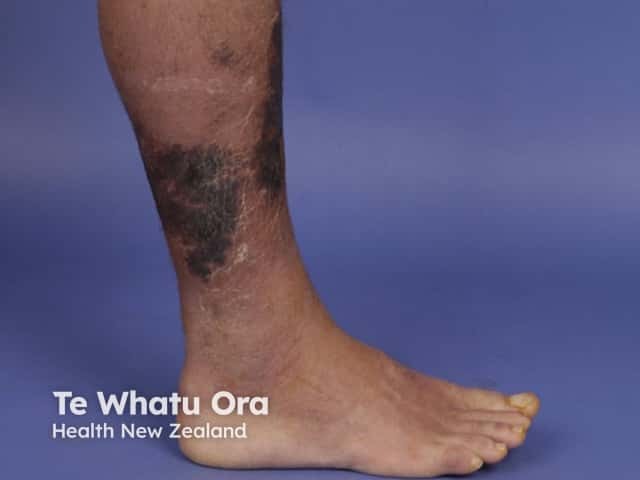
- The most common site of infection is the lower leg. Necrotising fasciitis may also affect upper limb, perineum, buttocks, trunk, head and neck.
- Symptoms appear usually within 24 hours of a minor injury.
- Pain is often very severe at presentation and worsens over time.
- There may be flu-like symptoms, such as nausea, fever, diarrhoea, dizziness and general malaise.
- Intense thirst develops as the body becomes dehydrated.
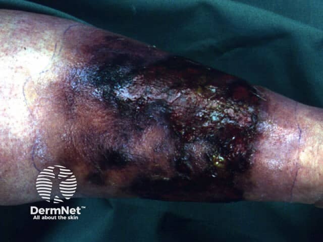
Clinical features after 3 to 4 days
- The affected area starts to swell and may show a purplish rash
- Large dark marks form that turn into blisters filled with dark fluid
- The wound starts to die and area becomes blackened (necrosis)
- Oedema is common
- A fine crackling sensation under the skin (crepitus) is due to gas in the tissues
- Severe pain continues until necrosis/gangrene destroys peripheral nerves when the pain subsides
- The infection may not improve when antibiotics are given
By about days 4–5, the patient is very ill with dangerously low blood pressure and high temperature. The infection has spread into the bloodstream and the body goes into toxic shock. The patient may have altered levels of consciousness.
Metastatic abscesses can develop in liver, lung, spleen, brain, pericardium, and rarely, in the skin.
See more images of necrotising fasciitis.
How is necrotising fasciitis diagnosed?
A thorough history and clinical examination are crucial in arriving at the diagnosis of necrotising fasciitis. Special care should be taken when examining immunocompromised patients, as the presentation of symptoms/signs may be atypical.
- Clostridial and streptococcal infections as a result of traumatic or surgical wound usually manifest quickly compared to necrotising fasciitis due to other organisms.
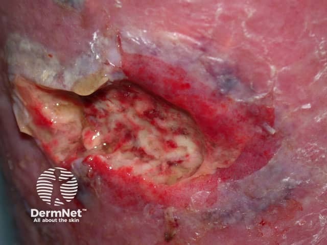
- A positive finger test is highly pathognomic for necrotising fasciitis. A 2–cm vertical incision is made in the affected skin and an index finger is pushed into the tissue. The test is positive if the finger passes through the subcutaneous tissue without resistance.
- There is poor adherence of tissue to the fascia on incising the site.
- Necrotic tissue/pus oozes out of the fascial planes.
- Dishwater-coloured fluid seeps out of the skin.
- Typically, necrotising fasciitis does not bleed.
Laboratory screening investigations
- White blood cell (WBC) count > 15.4 x 10⁹/L
- Serum sodium < 135 mmol/L
- Raised C-Reactive Protein (CRP) (> 16 mg/dL)
- Raised creatinine kinase (CK) level (> 600 U/L)
- Urea > 18 mg/dL.
Blood culture, deep tissue biopsy [see Necrotising fasciitis pathology], and Gram stain help in identifying the culprit organism(s) and guide the choice of antibiotic. If Staphylococcus aureus is detected, MRSA sensitivity test should be done. Blood cultures are usually negative for clostridial species.
Fungal culture should be performed in immunocompromised and trauma patients.
Imaging
X-ray, CT scan, and MRI identify areas of fluid collection, inflammation and gas within the soft tissues.
Laboratory risk indicator for necrotising fasciitis
The Laboratory Risk Indicator for Necrotising Fasciitis (LRINEC) is a tool that aids in distinguishing necrotising fasciitis from other tissue infections based on six parameters. A score of ≥ 6 favours necrotising fasciitis. This test is not appropriate for all cases and is not completely reliable.
LRINEC parameters
CRP (mg/dL) ≥15:
- 4 points
WBC count (109/L)
- <15: 0 points
- 15–25: 1 point
- >25: 2 points
Haemoglobin (g/L):
- >135: 0 points
- 110–135: 1 point
- <110: 2 points
Serum sodium (mmol/L) <135:
- 2 points
Creatinine (umol/L) >141:
- 2 points
Glucose (mmol/L) >10:
- 1 point
What is the treatment for necrotising fasciitis?
Once the diagnosis of necrotising fasciitis is confirmed, treatment should be initiated without delay.
- The patient must be hospitalised and is often admitted to an intensive care unit.
- The causative organism(s) should be identified and treated with high dose intravenous antibiotics. The initial antibiotic choice includes penicillin, clindamycin, metronidazole, cephalosporins, carbapenems, vancomycin, and linezolid. After the culture is reported, the choice is adjusted.
- It is absolutely vital that an experienced surgeon urgently removes all necrotic tissue (debridement).
- Supplemental oxygen, fluids, and medicines may be needed to raise blood pressure.
- Hyperbaric oxygen and intravenous immunoglobulin may also be considered.
Immediate surgical debridement improves survival and avoids complications of necrotising fasciitis. All infected tissue should be cleared away using adequate excision. Repeated debridements are carried out for a few days.
When the acute infection has subsided, the wound should be closed with skin grafting if required. Vacuum-assisted wound closing devices may be useful to heal a persistent ulcer.
What is the likely outcome?
Prompt diagnosis and treatment are essential to reduce the risk of death and disfigurement from necrotising fasciitis.
If diagnosed and treated early, most patients will survive necrotising fasciitis with minimal scarring. If there is significant tissue loss, later skin grafting will be necessary and in some patients amputation of limbs is required to prevent death.
Up to 25% of patients will die from necrotising fasciitis, due to complications such as renal failure and septicaemia (blood poisoning) and multiorgan failure.
References
- Machado NO. Necrotizing fasciitis: the importance of early diagnosis, prompt surgical debridement and adjuvant therapy. North Am J Med Sci. 2011; 3: 107–18. Journal
- Sadasivan J, Maroju NK, Balasubramaniam A. Necrotizing fasciitis. Indian J Plast Surg. 2013;46(3):472–8. doi: 10.4103/0970-0358.121978. Journal
- Puvanendran R, Huey JCM, Pasupathy S. Necrotizing fasciitis. Can Fam Physician. 2009;55(10):981–7. Journal
- Krieg A, Röhrborn A, Schulte Am Esch J, et al. Necrotizing fasciitis: microbiological characteristics and predictors of postoperative outcome. Eur J Med Res. 2009;14(1):30–6. doi:10.1186/2047-783x-14-1-30. Journal
- Misiakos EP, Bagias G, Patapis P, Sotiropoulos D, Kanavidis P, Machairas A. Current concepts in the management of necrotizing fasciitis. Front Surg. 2014;1:36. doi:10.3389/fsurg.2014.00036. Journal
- Cheng N-C, Tai H-C, Chang S-C, Chang C-H, Lai H-S. Necrotizing fasciitis in patients with diabetes mellitus: clinical characteristics and risk factors for mortality. BMC Infect Dis. 2015; 15: 417. doi: 10.1186/s12879-015-1144-0. Journal
- Hakkarainen TW, Kopari NM, Pham TN, Evans HL. Necrotizing soft tissue infections: review and current concepts in treatment, systems of care, and outcomes. Curr Probl Surg. 2014;51(8):344–62. doi: 10.1067/j.cpsurg.2014.06.001. PubMed Central
On DermNet
- Infectious gangrene
- Necrotising fasciitis — pathology
- Streptococcal skin infections
- Bacterial infections online course for health professionals
- Blistering skin conditions
- Australian box jellyfish stings
- Fournier gangrene
- Skin infections in people who inject drugs
Other websites
- Bacterial infections of the skin — DermNet e-lecture [Youtube]
- Blistering disorders — DermNet e-lecture [Youtube]
- Dermatologic manifestations of necrotizing fasciitis — Medscape