DermNet provides Google Translate, a free machine translation service. Note that this may not provide an exact translation in all languages

Translate

Panton-Valentine leukocidin Staphylococcus aureus

Last reviewed: May 2023

Author(s): Hyun Kyoung Lee, Medical Registrar; and Louise Reiche, Dermatologist, Palmerston North, NZ (2023)

Reviewing dermatologist: Dr Ian Coulson

Edited by the DermNet content department


toc-icon

What is Panton-Valentine leukocidin Staphylococcus aureus (PVL-SA)?

Panton-Valentine leukocidin (PVL) is an exotoxin produced by certain strains of Staphylococcus aureus (SA).

Staph. aureus with a virulence factor, such as the PVL exotoxin, enhances infection transmission and treatment resistance. PVL-SA is highly transmissible, lyses human neutrophils, and may cause recurrent skin and soft tissue infections (SSTIs) despite antibiotic treatment.

Who gets PVL-SA?

The disease prevalence of PVL-SA varies worldwide, for example 19% in Europe, 29% in the USA, 28–54% in Australia, and 47–75% in Western Africa. These figures may be an underestimate as PVL-SA is not routinely tested in laboratories unless specifically requested. 

Global dissemination has been enhanced through international travel. Patients with PVL-SA are often young adults with minimal exposure to healthcare settings.

The main risk factors for acquiring PVL SA, known as the ‘five Cs’ are:

  • Close contact (eg, contact sports, military recruits)
  • Contaminated items
  • Crowding
  • Cleanliness
  • Cuts and compromised skin integrity. 

What causes PVL-SA?

The Panton-Valentine leukocidin (PVL) toxin induces cytolytic pores in human polymorphonuclear neutrophils (PMNs), monocytes, and macrophages which in turn induces apoptosis. Two proteins comprise the PVL cytotoxin: LukS-PV and LukF-PV, encoded by their namesake genes. LukS-PV and LukF-PV are carried on bacteriophages. 

Cell death is toxin concentration dependent. Neutrophil apoptosis in the peripheral circulation weakens bacterial defence. This may explain the increased severity of necrotising pneumonia for PVL-positive patients, who have a higher frequency of neutropaenia.

Another contributing factor to the PVL toxin virulence is the upregulation of pro-inflammatory cytokines and nuclear factor-kappa B (NF-κB) in neutrophils, independently increasing necrotising infections.

What are the clinical features of PVL-SA? 

PVL-SA clinical presentations vary widely ranging from asymptomatic nasopharyngeal carriage, skin and soft tissue infections (SSTIs), to necrotising pneumonia. 

Cutaneous features

Distinctive features for PVL-positive (compared to PVL-negative) Staph. aureus include:

  • Recurrent boils and necrotising skin and soft tissue infections (SSTIs) in otherwise healthy patients
  • Recurrent SSTIs despite several courses of antibiotics
  • Pain or erythema disproportionate to the severity of clinical signs
  • Primary skin infections with no portal of entry, although disruption of the skin barrier can promote infection
  • Lesions that spread rapidly
  • High risk of transmission within households or close contacts.

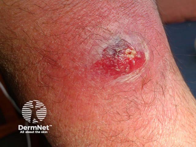
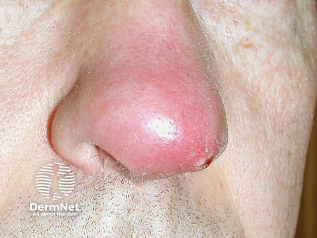
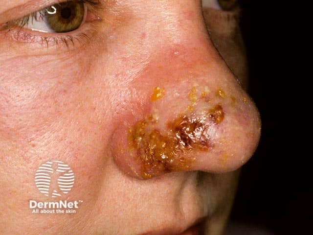
The most common PVL-SA SSTIs are:

Non-cutaneous features

PVL-SA can cause invasive infections in immunocompetent individuals, such as:

  • Necrotising pneumonia (often presents with flu-like symptoms and haemoptysis)
  • Musculoskeletal infections (eg, osteomyelitis; and, especially in children, pyomyositis or septic arthritis). 

What are the complications of PVL-SA?

  • Recurrent boils
  • Necrotising skin and soft tissue infections
  • Necrotising pneumonia (with a mortality rate up to 75%)
  • Osteomyelitis
  • Septic arthritis
  • Pyomyositis

How is PVL-SA diagnosed?

PVL-SA should be clinically suspected if there are recurrent abscesses/boils or necrotising SSTIs in an otherwise healthy individual, especially if a close contact is similarly affected.

Send bacterial swabs from the nose and infected areas of skin, and samples (eg, sputum, pus, or exudate) from other affected areas (as there may be coinfection) for microscopy, culture, and antibiotic sensitivities, specifically requesting PVL testing. 

The PVL virulence factor can be found in both methicillin-sensitive Staph. aureus, ie, MSSA strains (9–46%) and methicillin-resistant Staph. aureus ie, MRSA (74–100%). 

For cases of necrotising pneumonia, test for influenza due to the risk of coinfection.

What is the treatment for PVL-SA?

Consider prompt anti-staphylococcal regimen and an antitoxin agent; change to an appropriate antibiotic once susceptibilities are known.

  • Antitoxin agents include clindamycin, linezolid, and fusidic acid.
  • Note: subinhibitory concentrations of beta-lactam antibiotics can increase PVL production. 

Important hygiene measures for infected patients, close contacts, and carers include:

  • Separation of toiletries, and regular seperate washing of towels and clothes of infected persons
  • Frequent hand washing
  • Regular cleaning of shared household areas.

Specific measures

Specific treatment for PVL-SA depends on clinical disease severity at presentation.

  • Minor SSTIs: cover with dressings; consider topical antibiotics for 5 days
  • For moderate SSTIs: generally require 5–7 day oral antibiotics.
    • MSSA infections: clindamycin or flucloxacillin.
    • MRSA infections: clindamycin or rifampicin combined with either oral doxycycline, fusidic acid, or trimethoprim.
    • Incision and drainage may be required for abscesses.
  • For severe infections, empiric parenteral antibiotics for 48–74 hours until antimicrobial sensitivity for clindamycin is available.
    • Clindamycin or linezolid (if clindamycin-resistant).
    • If the patient deteriorates, consider intravenous immunoglobulin and early surgical debridement

How do you prevent PVL-SA?

PVL-SA preventative measures include hygiene and decolonisation measures for patients, close contacts, and those sharing the same household.

  • The hygiene measures listed in the treatment section also apply to prevention.
  • Repeated decolonisation 5-day course using:
    • Topical chlorhexidine 4% wash once daily (if allergic to chlorhexidine, use octenidine antimicrobial wash lotion)
    • Mupirocin nasal ointment three times daily (if mupirocin resistant, use neomycin-based nasal ointment).
  • After 5 courses of topical decolonisation, there is an 89% reduction in recurrence of PVL-SA infection.

What is the outcome for PVL-SA?

PVL-SA patients and close contacts who are treated early typically recover well. Clinician awareness, specific laboratory testing, and early appropriate decolonisation and antimicrobial use are key.

PVL-positive musculoskeletal infections generally have greater severity of inflammation and are more likely to require surgical intervention.

For necrotising pneumonia, the mortality rate in PVL-positive patients can be up to 75%, compared to 36% in PVL-negative cases.


Bibliography

  • Gillet Y, Issartel B, Vanhems P, et al. Association between Staphylococcus aureus strains carrying gene for Panton-Valentine leukocidin and highly lethal necrotising pneumonia in young immunocompetent patients. Lancet. 2002;359(9308):753–9. doi: 10.1016/S0140-6736(02)07877-7. Journal
  • Hanitsch LG, Krüger R, Hoppe P-A, et al. Outpatient decolonization after recurrent skin infection with Panton-Valentine leukocidin (PVL)-producing S. aureus: The importance of treatment repetition. PLoS One. 2020;15(4):e0231772. Journal
  • Hussain K, Bandyopadhyay A, Roberts N, et al. Panton–Valentine leucocidin‐producing Staphylococcus aureus: a clinical review. Clin Exp Dermatol. 2022;47(12):2150–8. Journal
  • Ritz N, Curtis N. The Role of Panton-Valentine Leukocidin in Staphylococcus aureus Musculoskeletal Infections in Children. Pediatr Infect Dis J. 2012;31(5):514–518. Journal
  • Saeed K, Gould I, Esposito S, et al. Panton–Valentine leukocidin-positive Staphylococcus aureus: a position statement from the International Society of Chemotherapy. Int J Antimicrob Agents. 2018;51(1):16–25. Journal

On DermNet

Books about skin diseases


Related information

Sign up to the newsletter