

Pityriasis alba — extra information
Introduction Demographics Causes Clinical features Diagnosis Treatment Prevention Outlook
What is pityriasis alba?
Pityriasis alba is a low-grade type of eczema/dermatitis mainly seen in children.
The name describes its appearance: pityriasis refers to the characteristic fine scale, and alba to its pale colour (hypopigmentation).
Who gets pityriasis alba?
Pityriasis alba is common worldwide with a prevalence in children of around 5%.
- Pityriasis alba mainly affects children and adolescents aged 3 to 16 years, but can occur in older and younger people.
- It affects boys and girls equally.
- The hypopigmentation of pityriasis alba is more prominent, and the condition perhaps more common, in dark skin compared to white skin.
What causes pityriasis alba?
The cause of pityriasis alba is unknown.
- Pityriasis alba often coexists with dry skin and atopic dermatitis.
- It often presents following sun exposure, perhaps because tanning of surrounding skin makes affected areas more prominent.
Researchers have not reached any conclusions about the relationship of pityriasis alba to the following:
- Ultraviolet radiation
- Excessive or inadequate bathing
- Low levels of serum copper and other trace elements
- Malassezia yeasts (which produce a metabolite, pityriacitrin, that inhibits tyrosinase thus causing hypopigmentation).
What are the clinical features of pityriasis alba?
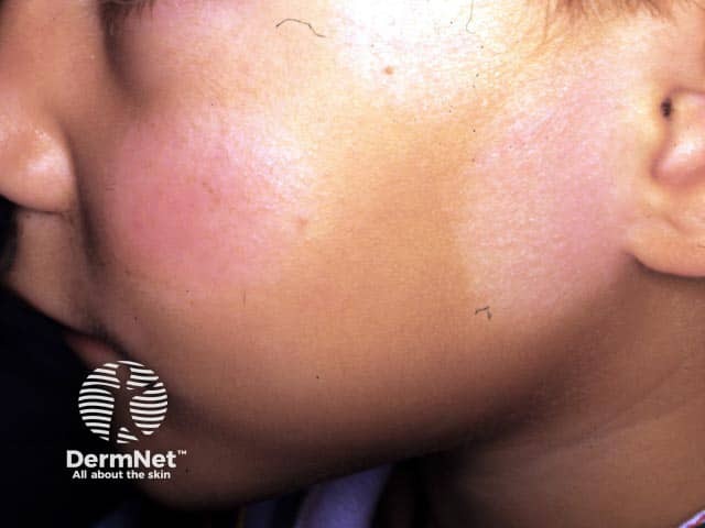
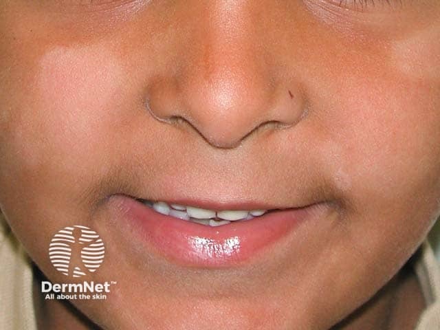
Classic pityriasis alba usually presents with 1 to 20 patches or thin plaques.
- Most lesions occur on the face, especially on the cheeks and chin.
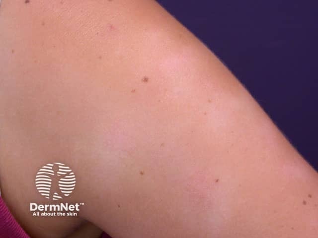
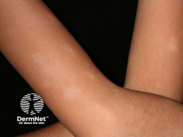
- Patches may also appear on the neck, shoulders, and upper arms and are uncommon elsewhere.
- Patch size varies from 0.5 to 5 cm in diameter.
- Patches are round, oval or irregular in shape.
- Pityriasis alba may have well-demarcated or, more usually, poorly defined edges.
- Itch is minimal or absent.
- Hypopigmentation is more noticeable in summer.
- Dryness and scaling is more noticeable in winter when environmental humidity tends to be lower.
Typically, a patch of pityriasis alba evolves through several stages.
- Slightly scaly pink patch or plaque with a just palpable papular surface.
- Hypopigmented patch or plaque with fine surface scale.
- Then post-inflammatory hypopigmented macule without scale.
- Resolution.
See more images of pityriasis alba ...
Dermoscopy of pityriasis alba
- Poorly defined pale area
- White structureless areas
- Faint pigment network
- Superficial white scale
- Normal hair colour
How is pityriasis alba diagnosed?
Pityriasis alba is usually a clinical diagnosis but may be confused with several other disorders that cause hypopigmentation.
To exclude these, investigations may include:
- Wood lamp examination: the hypopigmentation of pityriasis alba does not enhance, and there is no fluorescence
- Scrapings for mycology: microscopy and fungal culture are negative in pityriasis alba
- Skin biopsy: biopsy is rarely required, but may reveal mildly spongiotic dermatitis and reduction in melanin.
What is the treatment for pityriasis alba?
No treatment is necessary for asymptomatic pityriasis alba.
- A moisturising cream may improve the dry appearance.
- A mild topical steroid (0.5-1% hydrocortisone) may reduce redness and itch if present.
- Calcineurin inhibitors (pimecrolimus cream and tacrolimus ointment) may be as effective as hydrocortisone and have been reported to speed recovery of skin colour.
How can pityriasis alba be prevented?
The development or prominence of pityriasis alba can be reduced with sunscreen use to minimise sun tanning.
What is the outlook for pityriasis alba?
Pityriasis alba clears after an average of one year, with a range of a few months up to two or three years. The colour gradually returns completely to normal.
References
- Al-Refu K. Dermoscopy is a new diagnostic tool in diagnosis of common hypopigmented macular disease: A descriptive study. Dermatol Reports. 2018;11(1):7916. doi:10.4081/dr.2018.7916. PubMed
- Miazek N, Michalek I, Pawlowska-Kisiel M, Olszewska M, Rudnicka L. Pityriasis alba--common disease, enigmatic entity: up-to-date review of the literature. Pediatr Dermatol. 2015;32(6):786–91. doi:10.1111/pde.12683. PubMed
- Karanfilian KM, Behbahani S, Lambert MW, et al. The pathophysiology of pityriasis alba: time-dependent histologic changes. Clin Dermatol. 2020;38(3):354–6. doi:10.1016/j.clindermatol.2019.07.002. PubMed
- Jadotte YT, Janniger CK. Pityriasis alba revisited: perspectives on an enigmatic disorder of childhood. Cutis. 2011;87(2):66–72. PubMed
- Khafagy GM, Nada HR, Rashid LA, El-Samanoudy SI, Abd El-Sattar EM. Role of trace elements in pityriasis alba. J Trace Elem Med Biol. 2020;59:126422. doi:10.1016/j.jtemb.2019.126422. PubMed
- Ingram JR. Eczematous disorders. In: Griffiths C, Barker J, Bleiker T, Chalmers R, Creamer D (eds). Rook's Textbook of Dermatology [4 volumes], 9th edn, Wiley Blackwell, 2016:39.25–6.
On DermNet
Other websites
- Dermatologic Manifestations of Pityriasis Alba — Medscape Drugs & Diseases
- Pityriasis Alba — Medscape Drugs & Diseases
- Pityriasis alba — MedlinePlus