

Primary cutaneous follicle centre lymphoma — extra information
Introduction
Demographics
Causes
Clinical features
Complications
Diagnosis
Differential diagnoses
Treatment
Outcome
What is primary cutaneous follicle centre lymphoma?
Primary cutaneous follicle centre lymphoma (PCFCL) is a low-grade primary cutaneous B-cell lymphoma, defined as a lymphoma of germinal centre B-cells without extracutaneous involvement at the time of diagnosis.
Who gets primary cutaneous follicle centre lymphoma?
Primary cutaneous follicle centre lymphoma is the most common type of primary cutaneous B-cell lymphoma (PCBCL), accounting for 50%, and the second most common cutaneous lymphoma in the Western world. It accounts for 10–20% of all cutaneous lymphomas.
PCFCL mainly affects middle-aged white men (50–60 years of age). It is extremely rare in children.
What causes primary cutaneous follicle centre lymphoma?
Primary cutaneous follicle centre lymphoma has been associated with infections including Epstein-Barr virus, Helicobacter pylori, and Borrelia burgdorferi. It is a form of non-Hodgkin lymphoma.
What are the clinical features of primary cutaneous follicle centre lymphoma?
- Solitary or grouped plaques, nodules, or tumours
- Lesions are usually clustered in a localised area
- Pink, violaceous, skin-coloured, or erythematous in colour
- Lesions are usually not itchy and do not typically ulcerate
- Scalp, forehead, or trunk; rarely can be found on the lower limb
- Scalp lesions may present as a patch of hair loss and telangiectases
- Plaques and tumours on the back surrounded by macules and papules is a distinct variant called Crosti lymphoma or reticulohistiocytoma of the dorsum
- Miliary presentation of numerous scattered or clustered firm erythematous papules mainly on the forehead and cheeks
- Lesions can be disseminated in 10-20%
- Dermoscopy is non-specific
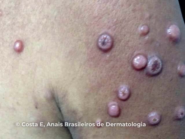
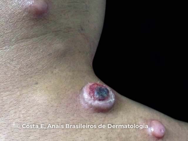
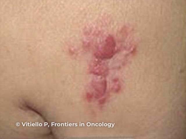
Figures 1&2: Costa EPW, Lucena BD, Amin GA, Bittencourt MJS, Dias LB Junior, Pires CAA. Primary cutaneous follicle center lymphoma. An Bras Dermatol. 2017;92(5):701–3. Figure 3: Vitiello P, Sica A, Ronchi A, Caccavale S, Franco R, Argenziano G. Primary cutaneous B-cell lymphomas: an update. Front Oncol. 2020;10:651.
What are the complications of primary cutaneous follicle centre lymphoma?
- Skin recurrence common after treatment — 30-40%
- Extracutaneous spread to lymph nodes or bone marrow — 5-10%
- Transformation to diffuse large B-cell lymphoma — rare
How is primary cutaneous follicle centre lymphoma diagnosed?
Primary cutaneous follicle centre lymphoma may be suspected clinically but is usually diagnosed on skin biopsy. Histology, immunohistochemistry, and molecular studies show:
- Dense dermal and subcutaneous proliferation of centrocytes (cleaved follicle centre cells) and centroblasts (large transformed cells) in a follicular and/or diffuse growth pattern, separated from the epidermis by a grenz zone
- Positive B-cell markers: CD19, CD20, CD22, CD79a, PAX5, and bcl-6 or CD10
- Negative bcl-2 protein, t(14;18) translocation, and immunoglobulin M expression.
Further investigations may include:
- Blood tests — full blood examination, metabolic panel, lactate dehydrogenase, C-reactive protein, protein electrophoresis, serology for B burgdorferi, HIV, and hepatitis
- Imaging — chest X-ray, PET/CT of chest, abdomen, and pelvis
- Bone marrow biopsy
- Lymph node biopsy if an enlarged node is detected on examination or imaging.
What is the differential diagnosis for primary cutaneous follicle centre lymphoma?
- Rosacea, acne, or folliculitis on the face
- Insect bites
- Other skin cancers including basal cell carcinoma, Merkel cell carcinoma, cutaneous T-cell lymphoma, secondary cutaneous lymphoma
- Histological differential diagnosis includes cutaneous lymphoid hyperplasia and primary cutaneous marginal zone lymphoma
What is the treatment for primary cutaneous follicle centre lymphoma?
- Solitary lesion or single site — surgical excision, radiotherapy
- Multiple lesions — radiotherapy, cryotherapy, intralesional steroids or rituximab
- Disseminated lesions — combination chemotherapy (R-CHOP), intravenous rituximab
Follow-up should be undertaken every 6 months including complete skin examination and lymph node palpation.
What is the outcome for primary cutaneous follicle centre lymphoma?
Primary cutaneous follicle centre lymphoma has an excellent prognosis. PCFCL may be stable, but lesions usually gradually enlarge if untreated and become locally aggressive. In rare cases, lesions may regress spontaneously.
The 5-year survival is >95%, or 40% if there is leg involvement.
Bibliography
- Costa EPW, Lucena BD, Amin GA, Bittencourt MJS, Dias LB Junior, Pires CAA. Primary cutaneous follicle center lymphoma. An Bras Dermatol. 2017;92(5):701–3. doi:10.1590/abd1806-4841.20175457. PubMed Central
- Massone C, Fink-Puches R, Laimer M, Rütten A, Vale E, Cerroni L. Miliary and agminated-type primary cutaneous follicle center lymphoma: report of 18 cases. J Am Acad Dermatol. 2011;65(4):749–55. doi:10.1016/j.jaad.2010.07.035. PubMed
- Skala SL, Hristov B, Hristov AC. Primary cutaneous follicle center lymphoma. Arch Pathol Lab Med. 2018;142(11):1313–21. doi:10.5858/arpa.2018-0215-RA. PubMed
- Vitiello P, Sica A, Ronchi A, Caccavale S, Franco R, Argenziano G. Primary cutaneous B-cell lymphomas: an update. Front Oncol. 2020;10:651. doi:10.3389/fonc.2020.00651. Journal
- Werner AC, Laver NV, Landmann DS. Primary cutaneous follicle center lymphoma of the eyelid: a case report and review of the literature in light of recent changes to the WHO classification of lymphoid neoplasms. Ocul Oncol Pathol. 2019;5(2):147-52. doi:10.1159/000491381. Journal
On DermNet
- Cutaneous B-cell lymphoma
- Cutaneous marginal zone lymphoma
- Cutaneous pseudolymphoma
- Cutaneous T-cell lymphoma
- Intravascular large B-cell lymphoma
- Lymphocytoma cutis
- Primary cutaneous diffuse large B-cell lymphoma pathology
Other websites
- Skin (cutaneous) B-cell lymphoma — Lymphoma Action
- Cutaneous B-cell lymphoma (CBCL) — Lymphoma Australia
- Cutaneous B-cell lymphoma — Lymphoma Research Foundation