Introduction
Uses
Contraindications
Classification
Procedure expectations
Benefits
Disadvantages
Side effects
Skin flaps are a type of surgical wound closure method that relocates tissue from one part of the body to another while preserving its intrinsic blood supply.
Unlike skin grafts, skin flaps are not reliant on the recipient site’s vascular bed to maintain tissue perfusion, thereby enhancing the viability of transplanted tissue.
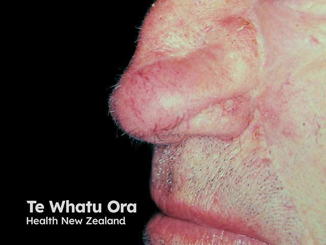
Skin flaps provide a means of restoring tissue continuity and function in areas where tissue has been lost or damaged eg, due to trauma, tumour resection, chronic wounds, or congenital defects.
Skin flaps are indicated when tissue defects cannot be safely or effectively closed by primary closure, healing by secondary intention, or skin grafting. Furthermore, flaps can incorporate various tissue types beyond just skin (eg, muscle, fascia, nerve, or bone) and meet specialised tissue needs such as bulk or sensation.
Contraindications to skin flap surgery depend on the flap type and the patient’s condition.
They may include the following:
As a precaution, poor patient health should be addressed or optimised before surgery:
Flap surgery is categorised using several classification systems. The tables below simplify these classifications into three main categories:
Classification by blood supply |
Description |
||||||||
|---|---|---|---|---|---|---|---|---|---|
Random pattern flaps |
Commonly used in local skin flaps, these flaps rely on blood supply from their network of small, unnamed vessels (subdermal plexuses). |
||||||||
Axial flaps |
Supplied by a named artery or group of arteries. These are preferred for covering larger or complex defects. |
||||||||
Classification by tissue type |
Description |
||||||||
|---|---|---|---|---|---|---|---|---|---|
Cutaneous (skin) flaps |
Consist of the epidermis, dermis, and subcutaneous fat, and are commonly used for surface-level coverage. |
||||||||
Fasciocutaneous flaps |
Incorporate fascia alongside the skin to add structural strength, which is especially valuable in areas under tension. |
||||||||
Myocutaneous flaps |
Contain muscle tissue and overlying skin, providing greater bulk and vascularisation, making it suitable for more extensive reconstruction. |
||||||||
Osseous flaps |
Include bone and are used for structural reconstruction and bony defects eg, maxillofacial defects. |
||||||||
Composite flaps |
Consist of more than one tissue type (eg, skin, fascia, muscle, bone, cartilage) to repair complex defects. |
||||||||
Classification by donor site |
Description |
||||||||
|---|---|---|---|---|---|---|---|---|---|
Local flaps |
The donor and recipient sites are adjacent. There are four types of movement for local flaps:
|
||||||||
Distant flaps |
The donor and recipient sites are far apart. These can be pedicled or free:
|
||||||||
The dermatologist or surgeon will discuss the indication for a skin flap, the procedure involved, and common complications.
Patients should inform their doctor of any:
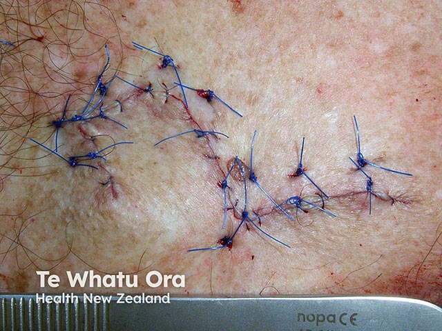
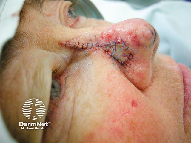
Most local skin flap surgeries are performed under local anaesthesia. The procedure may create two wound sites: one at the recipient site and one at the donor site. In local flaps, these sites are typically adjacent and can be dressed under a single bandage.
The wound may be tender for 1-2 hours post-excision when the local anaesthetic wears off. Stitches are typically removed 5-10 days after the operation.
A slight amount of pinkness and tenderness around the wound edges is normal. However, increasing redness, pain, and swelling of the wound, along with fever or a general feeling of unwellness, can indicate an infection that requires antibiotics.

