DermNet provides Google Translate, a free machine translation service. Note that this may not provide an exact translation in all languages

Translate

Trichoscopy of scalp infestations

May 2022

Author: Dr Ahmed Sadek, Cairo Hospital for Dermatology & Venereology (Al-Haud Al-Marsoud), Egypt. May 2022.

Contributors: Dr Dalia Hossam, Dr Radwa Magdy, Dr Nehal Saied, Dr Noha Hashem, Dr Safaa Yehia Negm, Dr Moshera Saied El Bahrawy, Dr Amira Ragab, Dr Amal Wagih, Dr Haidy El-Hussieny, Dr Mona Ragib, Dr Hala Amer. Copy edited by Gus Mitchell. May 2022


toc-icon
Table of contents
arrow-right-small

Pediculosis of the scalp

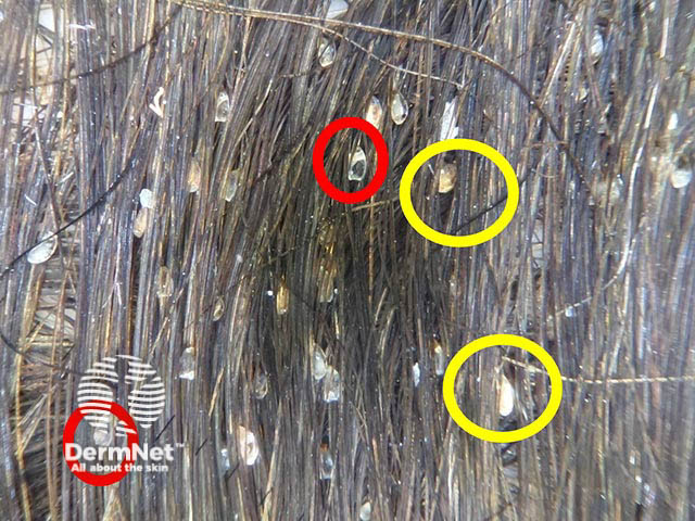
Pediculosis capitis (head lice) occurs with an infestation of the scalp hair with the human head louse (Pediculus humanus capitis), an external obligate parasite that inhabits the scalp and feeds by sucking blood. Any age may be affected, however it is most prevalent among schoolchildren.

Trichoscopic features and uses in pediculosis

  • Identification of the mature insect and its eggs in the scalp of the infected objects. 
  • Assess if the parasites are dead or nits are empty.
  • Viable nits containing nymphs appear as dark brown pyriform-shaped structures of 0.8 mm in length, attached to the shaft; hatched nits are translucent and typically show a plane and fissured free ending and they may persist after the eradication of live head lice.
  • Assessment of the efficacy of treatment and follow up of patients.
  • Differentiation between nits and pseudo-nits (hair casts).
  • Screening of family members, thus reducing the risk of further infestation. 

Trichoscopy of scalp infestations

Scabies of the scalp

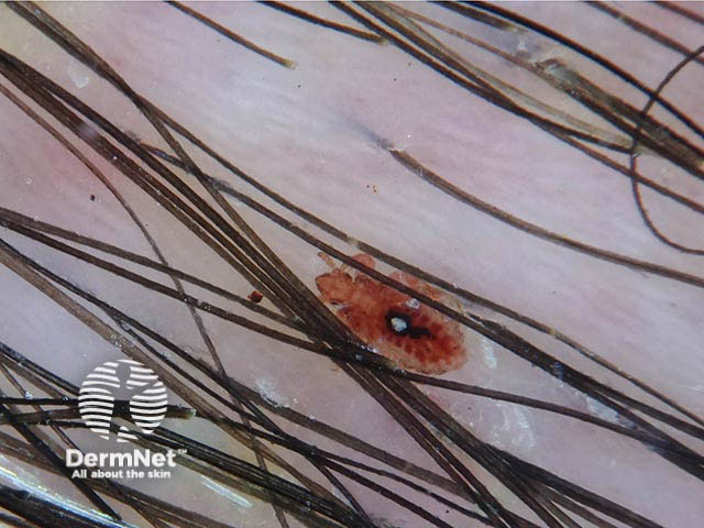
Scabies is a parasitic infestation caused by the mite Sarcoptes scabiei var hominis. It usually only affects the scalp of the newborn and those that are immunocompromised, showing the Norwegian or crusted form of scabies where the face may also be affected. Scabies infestation produces diagnostic burrows; the tunnel dug by the female Sarcoptes mite, in the host’s epidermis, to lay her eggs.

Trichoscopic features and uses in scabies

  • Identification of burrows and mites. They appear as a thin track of white scales with a small dark brown triangular-shaped structure representing the anterior body of the mite ("delta-wing jet with contrail sign")
  • "Hang glider sign”, a brown triangle corresponding to the anterior part of the mite.
  • Burrows filled with eggs may appear as a “string of pearls”.
  • Monitor the patient's response to therapy using dermoscopy guided skin scraping.
    • Blurry indistinct mites are a sign of successful therapy.

Other features

These features are best seen with higher magnifications:

  • The "noodle sign" in Norwegian scabies: accumulation of hundreds of burrows in the same dermoscopic field
  • Cutaneous nodules as described in neonatal scabies.


Bibliography

  • Badri T, Hammami H, Benmously R, Mokhtar I, Fenniche S. Dermoscopic diagnosis of pediculosis capitis. Acta Dermatovenerol Alp Pannonica Adriat. 2010;19(3):45–6. Journal
  • Bollea Garlatti LA, Torre AC, Bollea Garlatti ML, Galimberti RL, Argenziano G. Dermoscopy aids the diagnosis of crusted scabies in an erythrodermic patient. J Am Acad Dermatol. 2015;73(3):e93–e95. doi:10.1016/j.jaad.2015.04.061. Journal
  • Micali G, Tedeschi A, West DP, Dinotta F, Lacarrubba F. The use of videodermatoscopy to monitor treatment of scabies and pediculosis. J Dermatolog Treat. 2011;22(3):133–7. doi:10.3109/09546631003649687. Journal
  • Piccolo V. Update on Dermoscopy and Infectious Skin Diseases. Dermatol Pract Concept. 2019;10(1):e2020003. Published 2019 Dec 31. doi:10.5826/dpc.1001a03. Journal
  • Zalaudek I, Argenziano G. Images in clinical medicine. Dermoscopy of nits and pseudonits. N Engl J Med. 2012;367(18):1741. doi:10.1056/NEJMicm1103059. Journal

On DermNet

Books about skin diseases


Related information

Sign up to the newsletter