Introduction
Alopecia areata
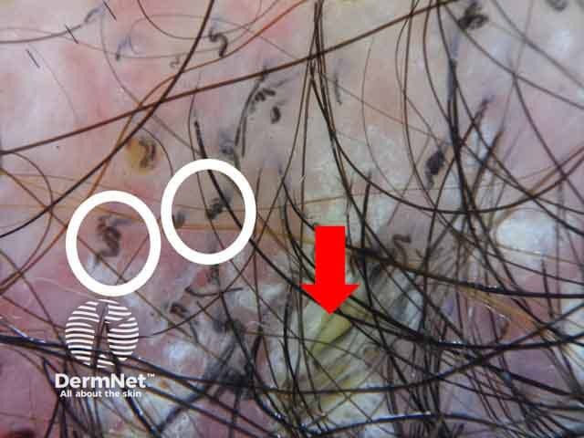
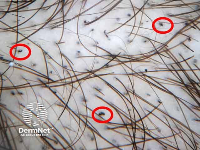
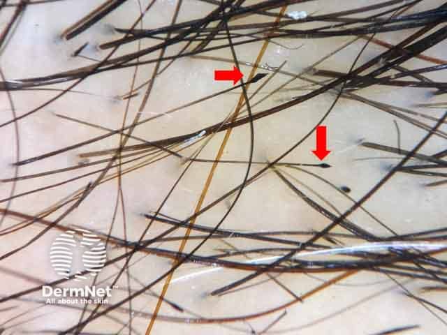
Tinea capitis
Trichotillomania
Traction alopecia
Temporal triangular alopecia
Many scalp and hair disorders present with focal hairless patches which require meticulous examination to differentiate between cicatricial (scaring) and noncicatricial (non-scarring) alopecia and to identify their exact cause. Trichoscopy can be used as a non-invasive tool for rapid diagnosis of different types of alopecia.
Main causes of localised noncicatricial alopecia:
Refer to generalised noncicatricial hair loss for further information on the trichoscopy of alopecia areata.
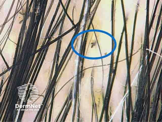
Tinea capitis is a superficial fungal infection of the scalp commonly affecting children, however it may present in immunosuppressed adults. The disease is primarily caused by dermatophytes, Trichophyton and Microsporum genera, that cause endothrix or ectothrix type hair shaft infection respectively.
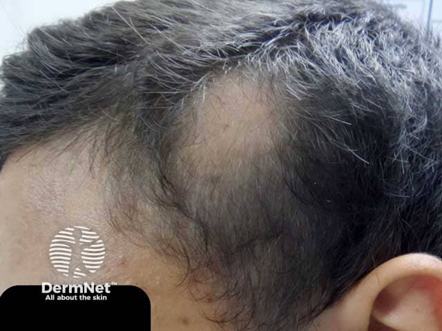
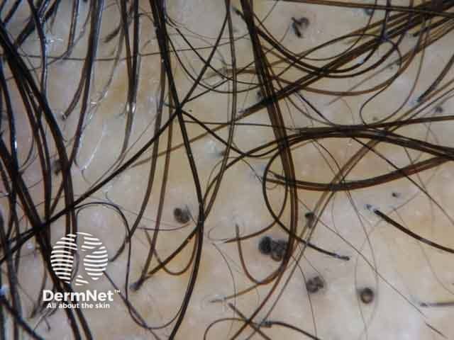
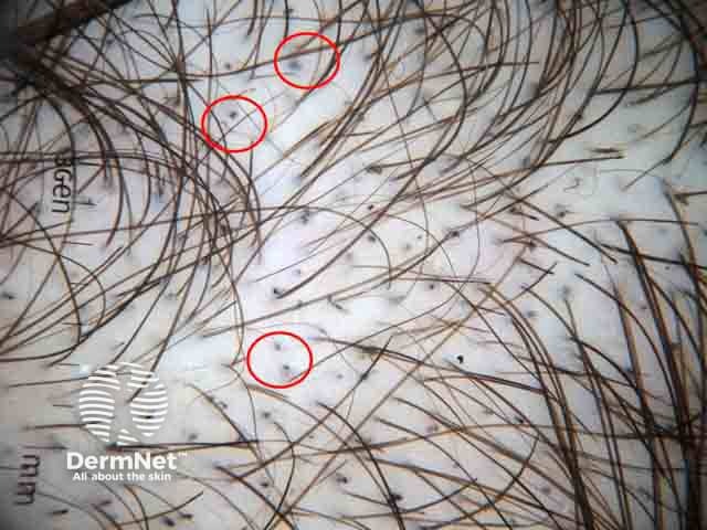
The clinical presentation is typically of a single or multiple patches of hair loss, sometimes with a black dot pattern, that may be accompanied by inflammation, scaling, pustules, and itching.
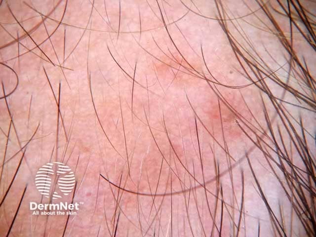
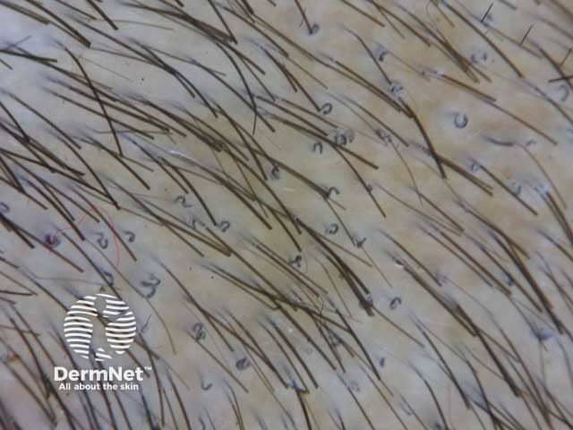
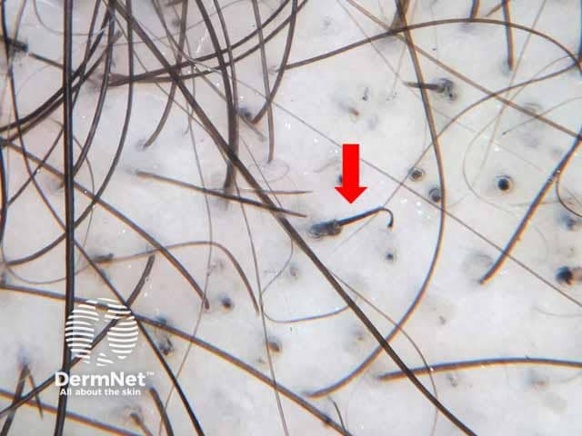
Specific features:
Comma and corkscrew hairs represent the bending of the affected hair shafts due to the invasion with fungal hyphae.

Other features:
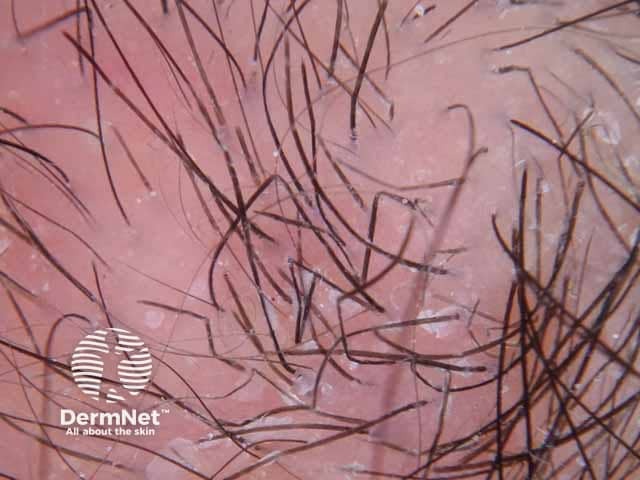
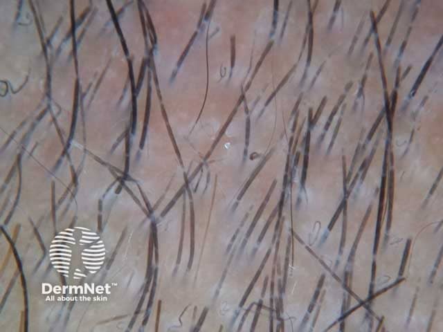
Trichotillomania is a body-focused repetitive behaviour disorder. Sufferers may derive pleasure, gratification, or relief when pulling out their hair. It presents with irregular patches of alopecia, with hairs of variable length commonly affecting the vertex or parietal scalp.
Specific features:
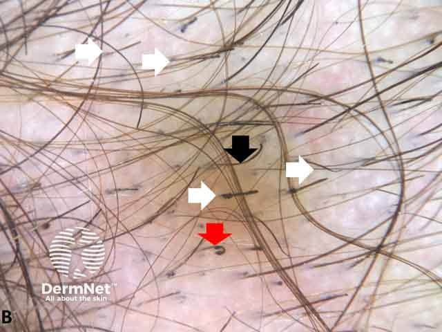
Other features:
Refer to trichoscopy of generalised noncicatricial hair loss for further information on the trichoscopy of tractional alopecia.
Temporal triangular alopecia is a non-scarring, circumscribed alopecia is often located unilaterally in the frontotemporal region during early childhood and remains stationary throughout life.