Main menu
Common skin conditions

NEWS
Join DermNet PRO
Read more
Quick links
Author: Dr Delwyn Dyall-Smith, Dermatologist, Wagga Wagga, NSW, Australia. DermNet Editor in Chief: Adjunct A/Prof Amanda Oakley, Dermatologist, Hamilton, New Zealand. Copy edited by Gus Mitchell/Maria McGivern. August 2018.
Introduction
Causes
Demographics
Prevalence
Symptoms and signs
Diagnosis
Treatment
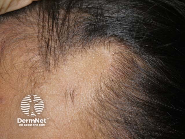
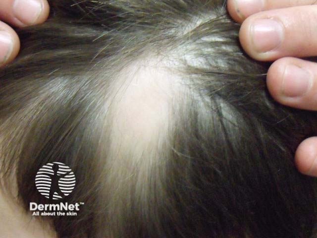
Temporal triangular alopecia, also known as congenital triangular alopecia, is a non-scarring form of hair loss usually seen on the frontotemporal scalp.


The cause of temporal triangular alopecia is unknown. It is usually sporadic but has been reported to run in families, suggesting a possible genetic link. Temporal triangular alopecia has been reported in a number of genetic conditions, including Down syndrome and phacomatosis pigmentovascularis.
Temporal triangular alopecia most commonly presents in children aged 2–9 years, although it can be present at birth or may first appear in adult life. It affects both men and women and is more commonly seen in light-skinned people.
A survey of 6200 patients attending a dermatology clinic found the incidence of temporal triangular alopecia to be 0.11%. However, this does not necessarily reflect the general population. Many believe the condition is under-reported due to affected individuals not presenting with symptoms. Misdiagnosis of temporal triangular alopecia as alopecia areata, male pattern alopecia, traction alopecia, trichotillomania, or congenital aplasia cutis would also contribute to the impression of rarity.
Temporal triangular alopecia appears as a triangular or spear-shaped loss of hair, with the ‘point’ of the triangle directed up and back. The shape is sometimes round or oval. It usually does not cause any symptoms, but sometimes patients report dysaesthesia in the lesion.
The lesion most commonly appears on the temporal scalp on one side only, although can affect both sides. Involvement of the occipital hairline has also been reported.
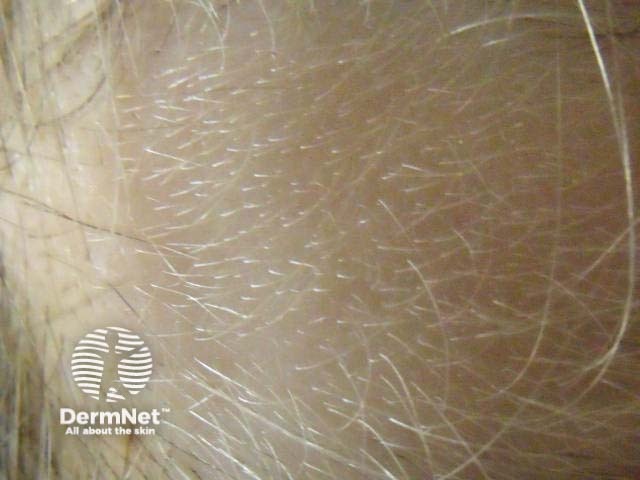
Temporal triangular alopecia often has a fringe of terminal hairs (mature adult hair) along the frontal hairline and sometimes also a tuft of hair within the lesion. Vellus hairs (soft finer hair) are seen throughout the area.
The lesion remains unchanged throughout life. Despite the presence of vellus hairs, there is no hair regrowth. There are no clinical signs of inflammation or scarring.
The diagnosis of triangular temporal alopecia is usually clinical.
Dermoscopy can distinguish temporal triangular alopecia from alopecia areata and male pattern alopecia. The dermoscopic features to look for are the presence of vellus hairs throughout the lesion with terminal hairs at the edge. There is no inflammation or scarring. The ‘exclamation mark’ hairs (short broken off hairs that resemble an exclamation mark), yellow dots, and black dots of alopecia areata are not present, nor are the 'streamers' (residual fibrovascular tracts) of male pattern alopecia.
A skin biopsy is not usually required, but histology shows a normal number of hair follicles in the superficial dermis, mainly of the vellus type.
Suggested criteria for diagnosis include:
Treatment is generally not required and is mostly ineffective.
Surgical excision of the lesion, if the lesion is small, or hair transplants, if cosmetically significant, have been used successfully.