DermNet provides Google Translate, a free machine translation service. Note that this may not provide an exact translation in all languages

Translate

Verrucous haemangioma pathology

Author: Adjunct A/Prof Patrick Emanuel, Dermatopathologist, Clínica Ricardo Palma, Lima, Peru. DermNet NZ Editor in Chief: Adjunct A/Prof Amanda Oakley. Copy edited by Gus Mitchell. September 2018.


toc-icon
Table of contents
arrow-right-small

Introduction

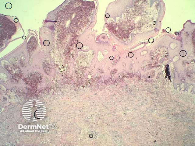
Verrucous haemangioma (American spelling hemangioma) presents as blue-red, vascular papules, plaques, or nodules, which later become warty in appearance. These lesions do not resolve spontaneously and have a tendency to recur after excision if margins are inadequate.

Histology of verrucous haemangioma

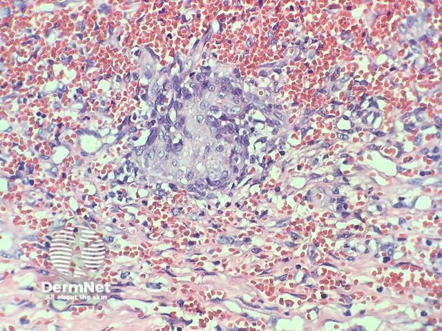
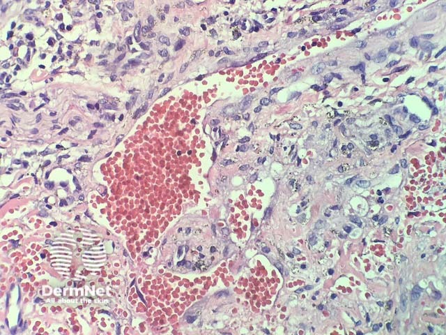
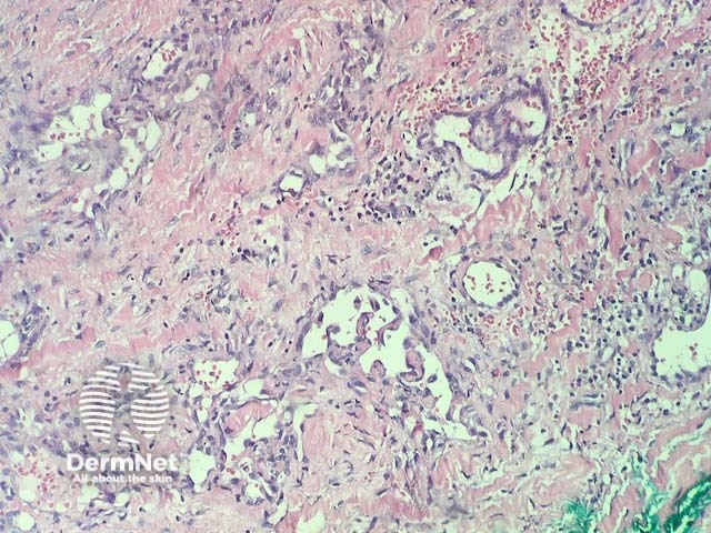
In verrucous haemangioma, the histopathology shows irregular papillomatosis, acanthosis and hyperkeratosis of the epidermis. The dermis shows multiple, thin-walled, dilated blood-filled spaces. Intravascular thrombosis with recanalisation and haemorrhage can be seen (figures 1–5).

Verrucous haemangioma pathology

Special studies for verrucous haemangoma

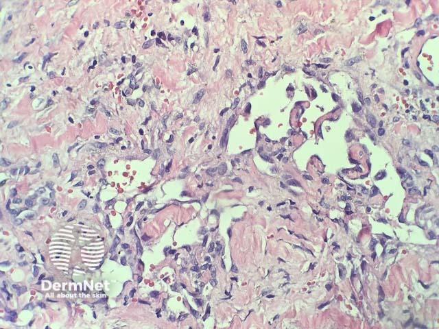
Vascular markers can highlight the extent of the lesion (CD31, CD34).

Differential diagnosis for verrucous haemangioma

Other conditions that should be considered include:

  • Angiokeratoma — these are generally smaller and are superficial. Involvement of the deep dermis and subcutis is not a feature of angiokeratoma
  • Kaposi sarcoma — some areas of verrucous haemangioma can have infiltrative growth and mimic Kaposi sarcoma. Immunohistochemistry with HHV8 is negative in verrucous haemangioma.


References

On DermNet

Books about skin diseases


Related information

Sign up to the newsletter