Created 2008.
The dermoscopy 3-point checklist for early detection of skin cancer is fairly easy to learn and has a high sensitivity for melanoma. (1) There a high likelihood of malignancy (melanoma or basal cell carcinoma) if a pigmented skin lesion has any two of these criteria. The 3-point checklist has been designed to allow non-experts not to miss detection of melanomas. However, it is not as specific as pattern analysis (described in a later section).
The main aim of the 3-point checklist is to determine whether the lesion being examined should undergo a biopsy. It does not an require accurate diagnosis to be made as the finer features of the lesion are not examined.
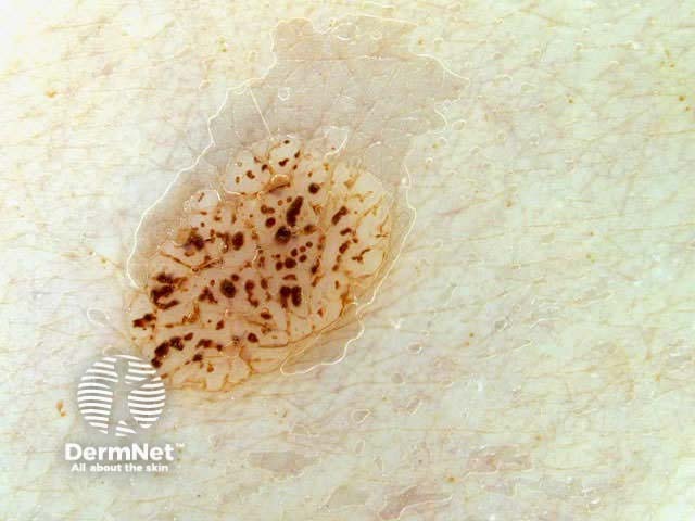
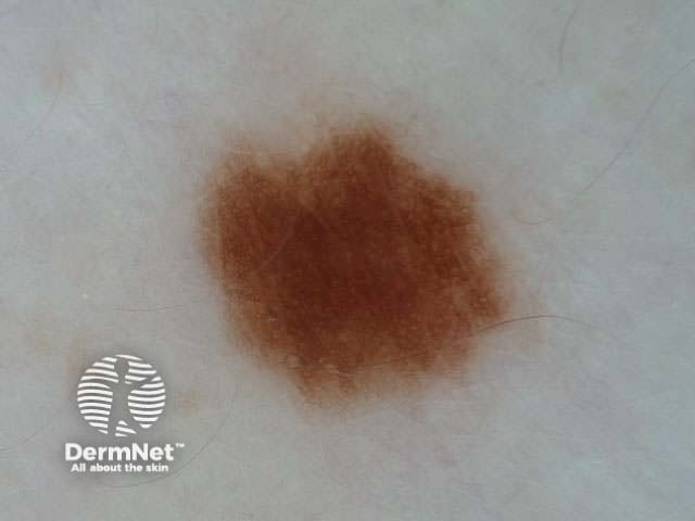
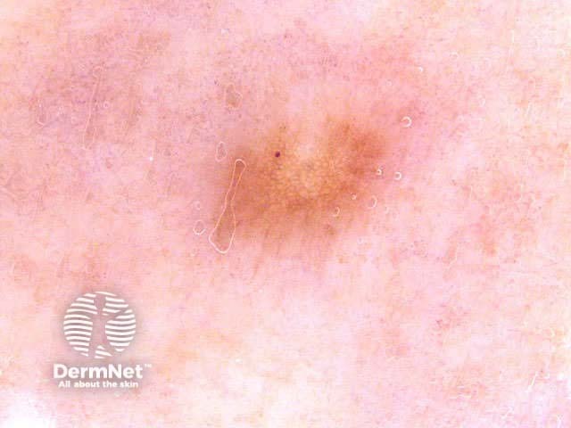
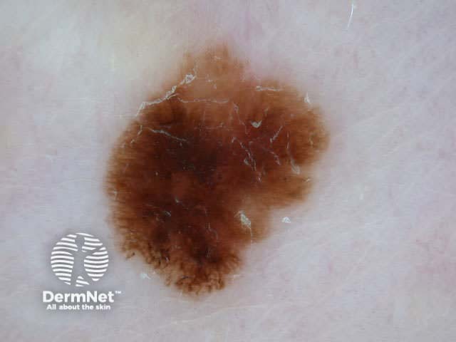
The following lesions demonstrate approximate symmetry of colour and structure. Shape is not considered.
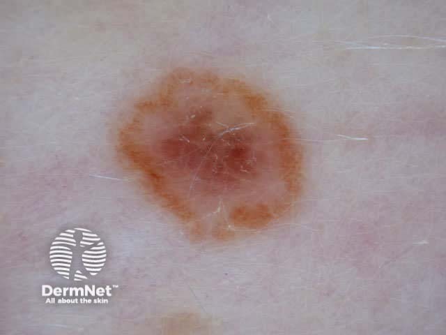
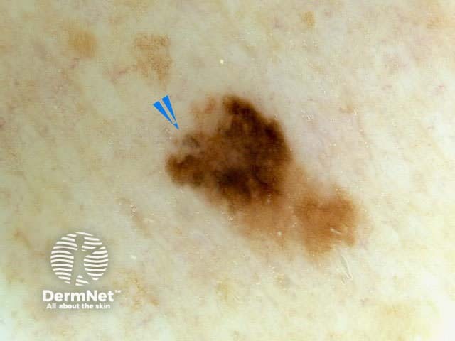
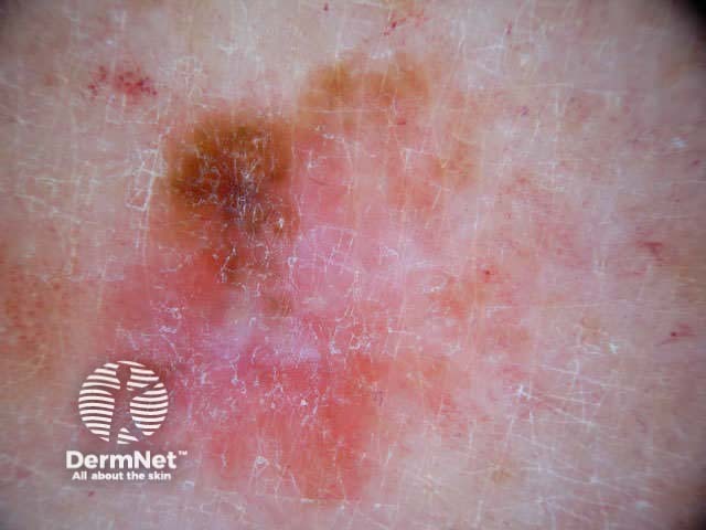
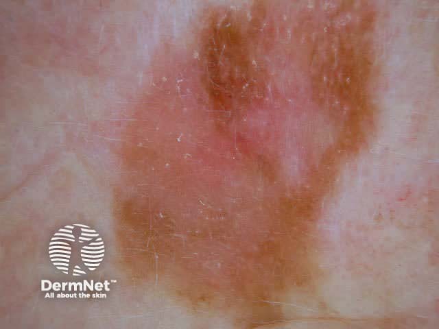
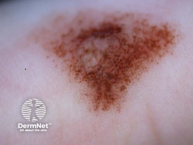
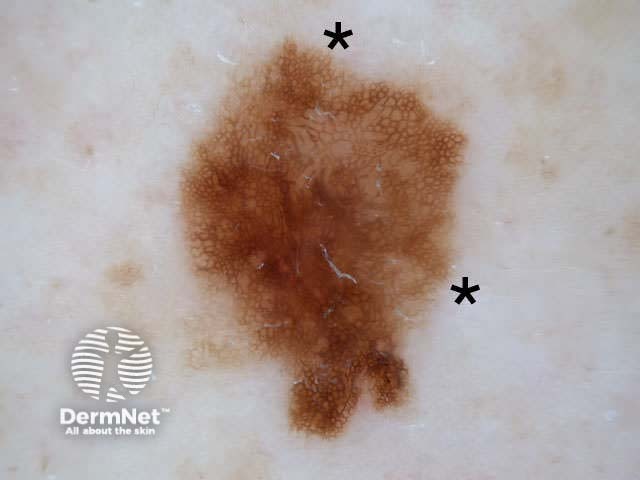
The lesions below demonstrate asymmetry of colour or structure in one or two axes. Not all asymmetrical lesions are malignant.
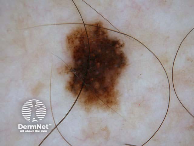
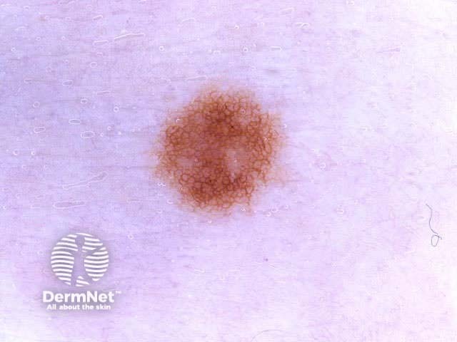
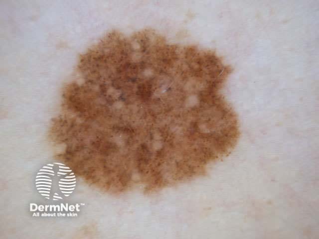
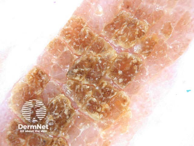
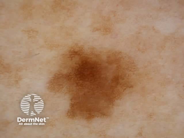
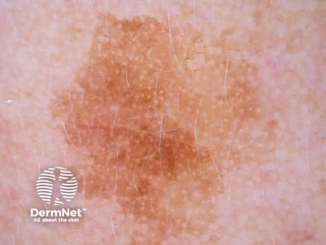
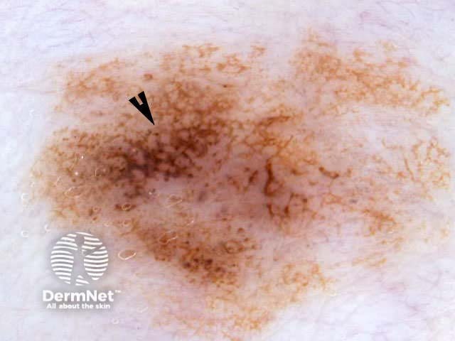
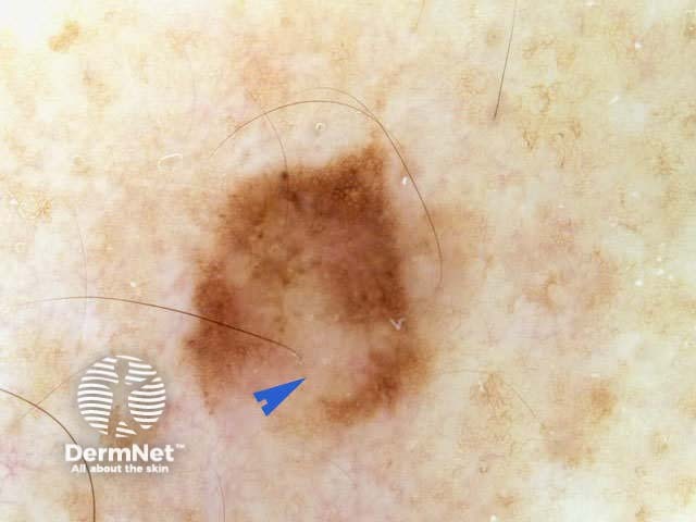
The following lesions are considered to have typical pigment network.
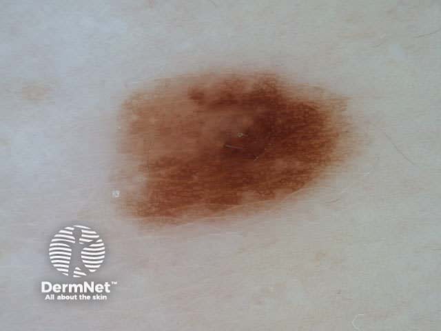
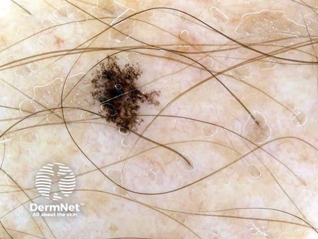
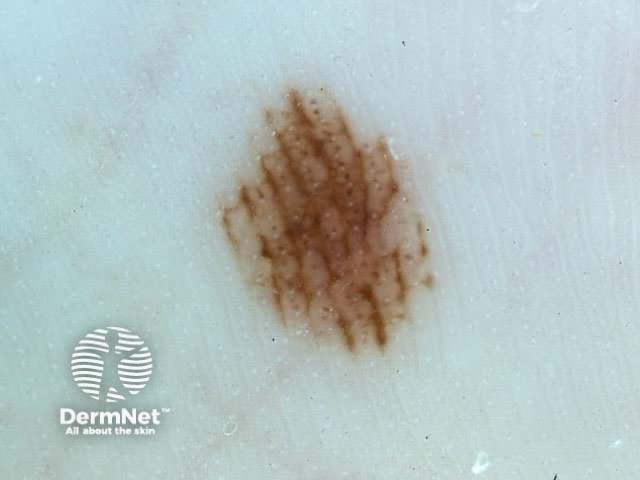
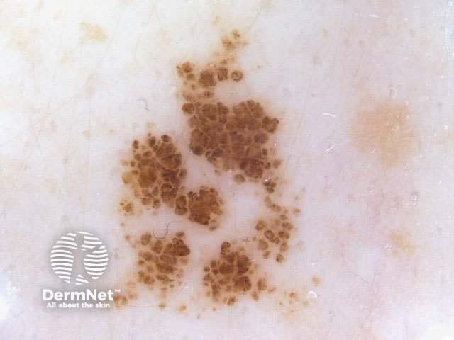
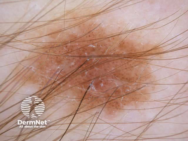
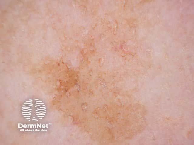
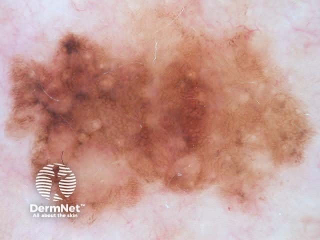
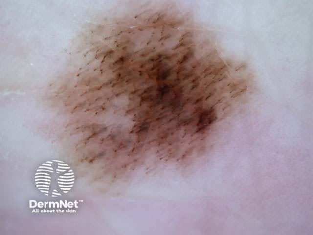
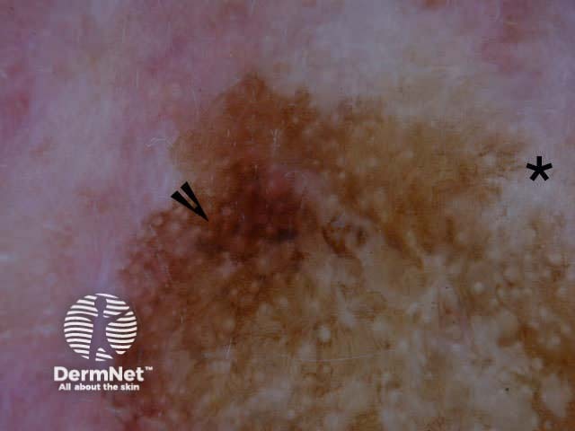
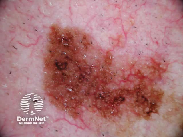
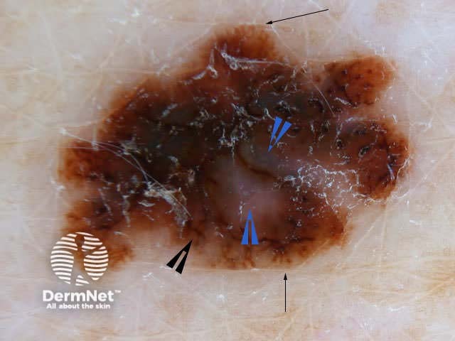
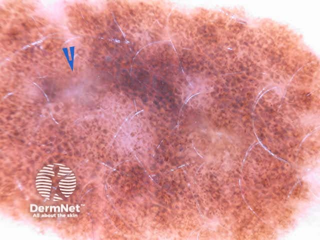
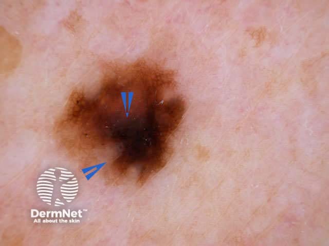
The lesions below have atypical pigment network, with irregular holes and thick lines (broadened network). Streaming or pseudopods would also be considered atypical. Not all lesions with atypical network prove to be malignant.
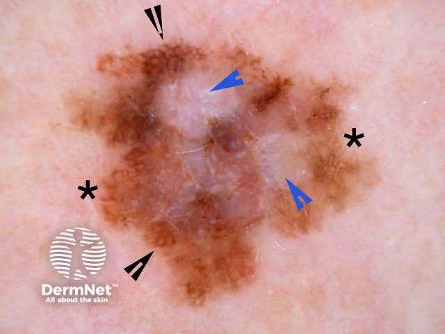
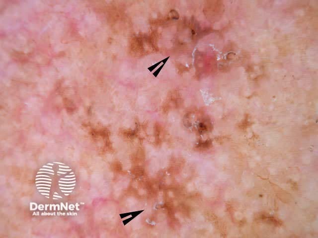
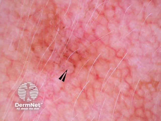
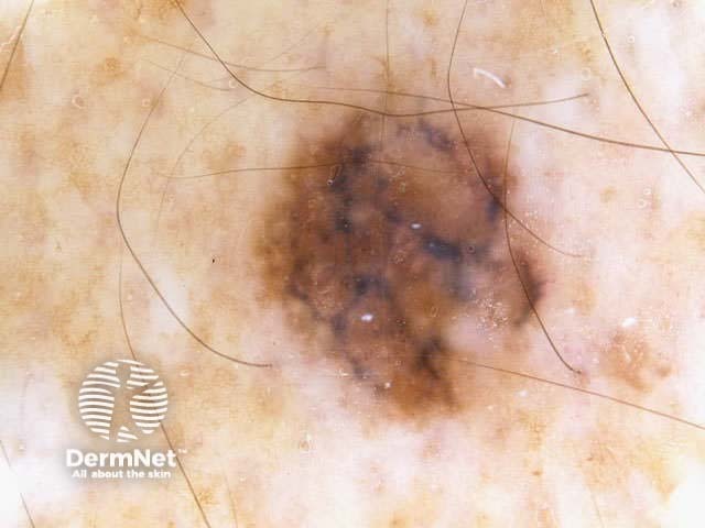
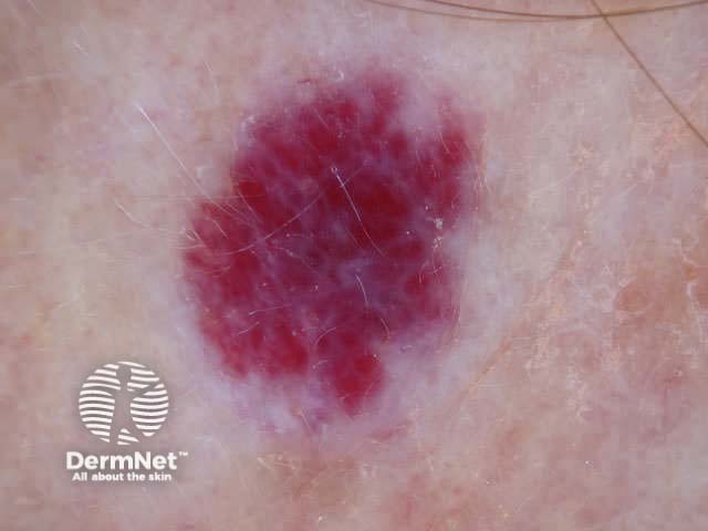
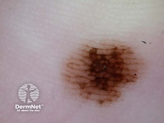
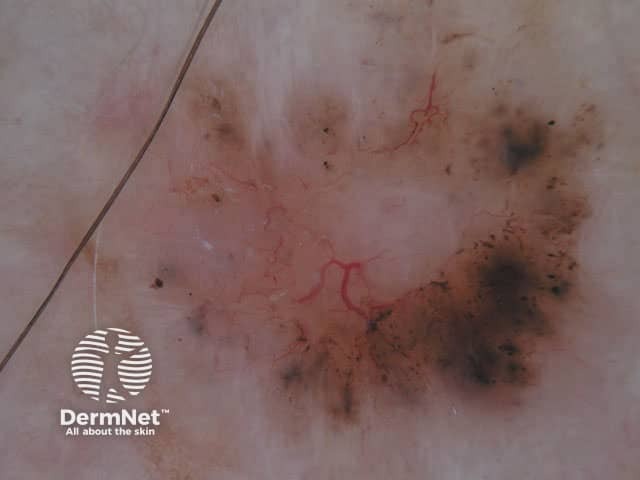
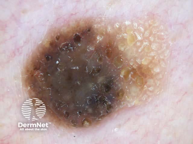
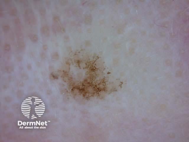
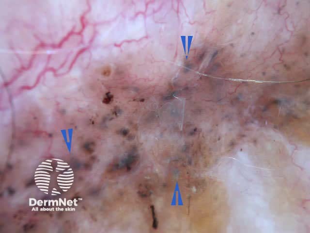
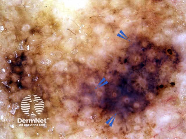
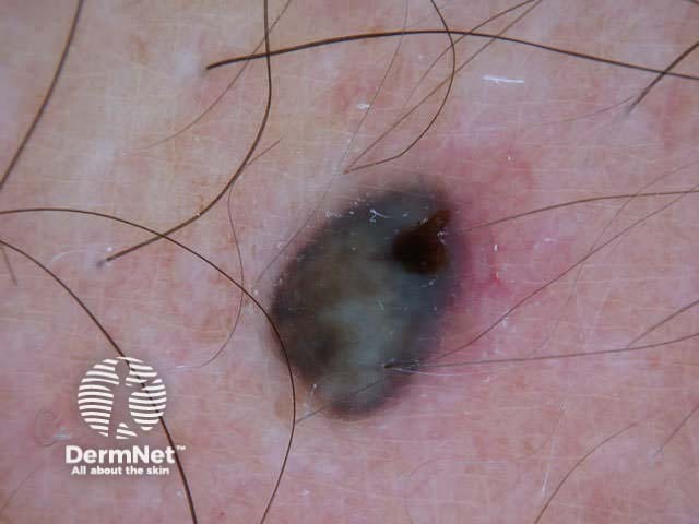
Blue-white structures can refer to any type of blue and/or white colour, i.e. combination of blue-white veil and regression structures, as shown in the following pictures. The colour can be subtle. Not all lesions with blue-white structures are malignant.
Practice dermoscopy, evaluating skin lesions using the 3-point checklist.
See the DermNet bookstore.

