Created 2008.
In dermoscopy, the first step algorithm identifies whether a lesion is melanocytic or nonmelanocytic. Pattern analysis is the method preferred by many expert dermoscopists to diagnose melanocytic lesions and to differentiate benign melanocytic lesions from malignant melanoma. Pattern analysis refers to the simultaneous assessment of the diagnostic value of all dermoscopy features shown by the lesion.
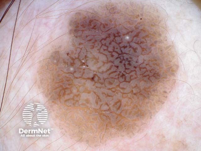
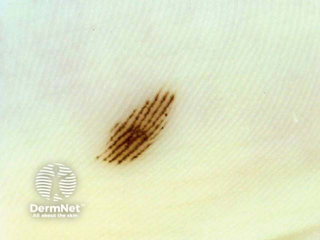
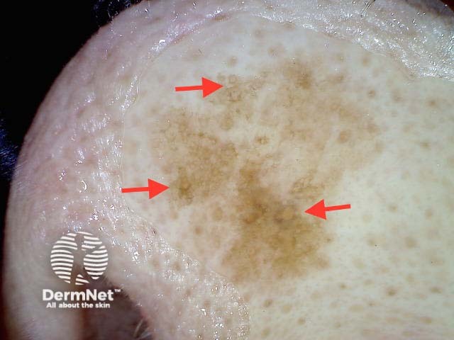
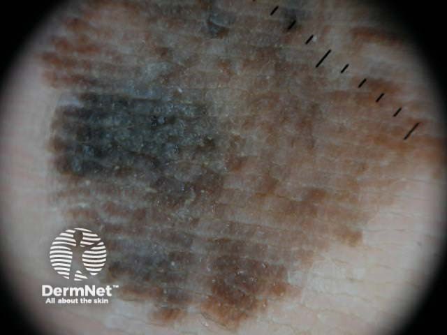
In general terms, benign lesions have few colours, a regular structure and are symmetrical in pattern. Malignant tumours often have several colours (especially melanoma), disordered structure and asymmetry of pattern.
If conventional or morphological pattern analysis appears too complicated, use modified pattern analysis, or the 3-point checklist to identify malignant pigmented lesions.
Non-concentric multicomponent pattern is highly suspicious of malignancy, but is occasionally seen in benign lesions, eg collision tumours, recurrent naevus, congenital melanocytic naevus.

Modified or revised pattern analysis is the descriptive system described by Kittler et al, now widely adopted. Terminology is much simpler in this system than in tranditional metaphoric dermoscopic pattern recognition. It is a single step system. Describe the lesion and decide if it should be excised or not. Lesions with asymmetry of structural elements and patterns are suspicious of malignancy.
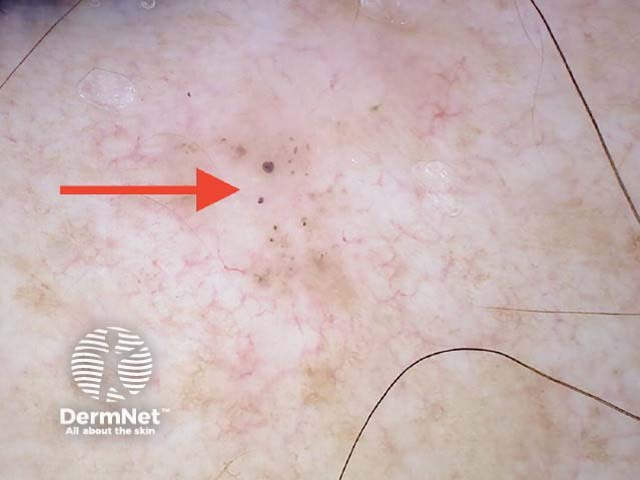
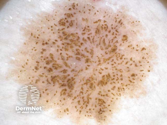
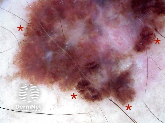
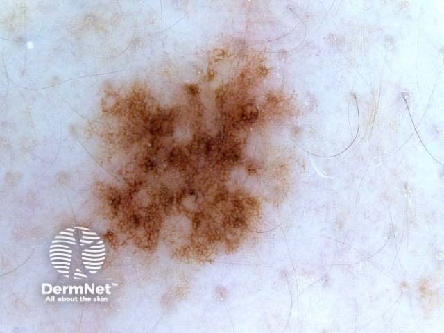
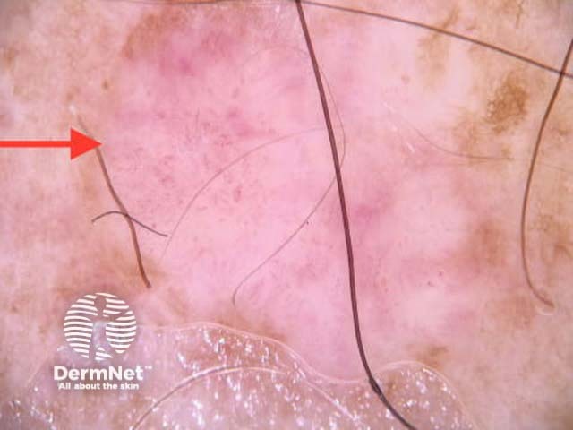
Dermoscopic features are broken down into elements: lines and rounded structures. The absence of a defined structure is described as structureless.
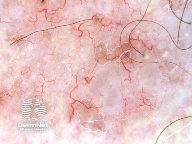
Global dermoscopic patterns for melanocytic lesions are described using these terms.
Non-melanocytic lesions are made up of the same elements.
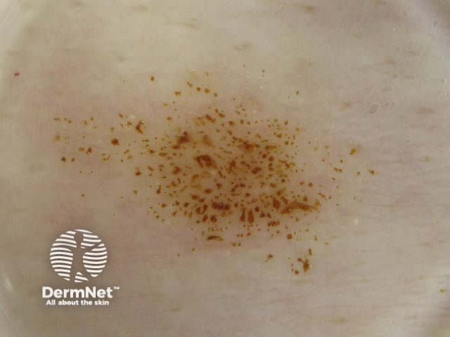
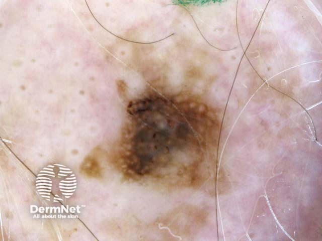
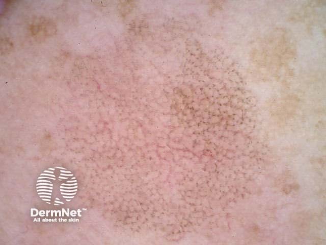
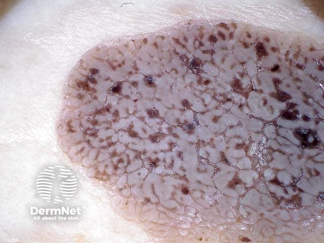
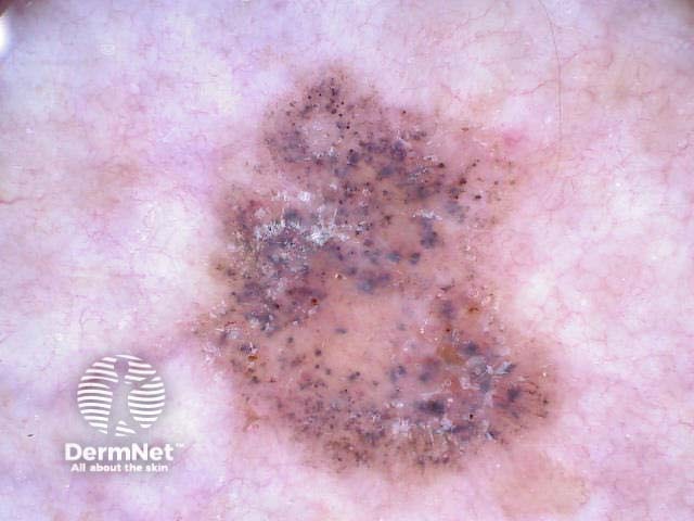
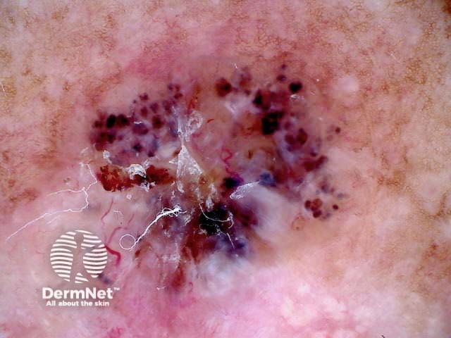
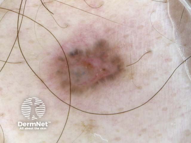
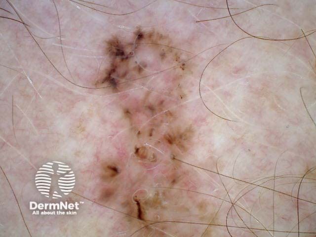
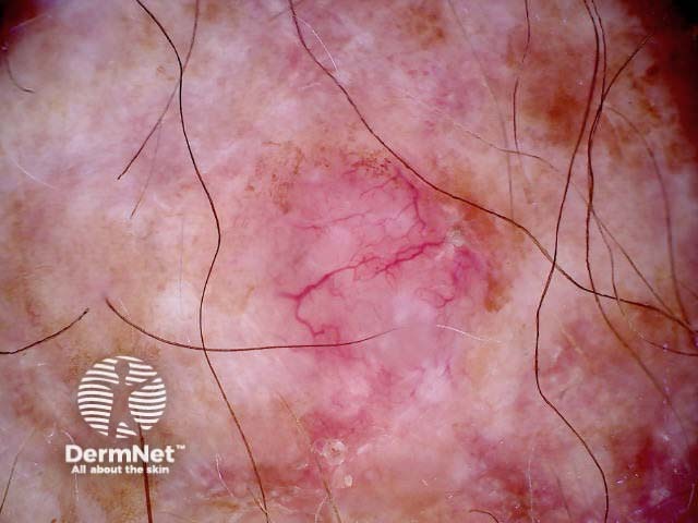
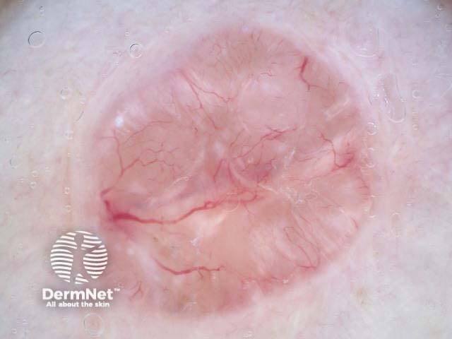

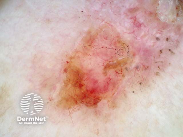
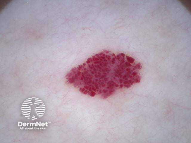
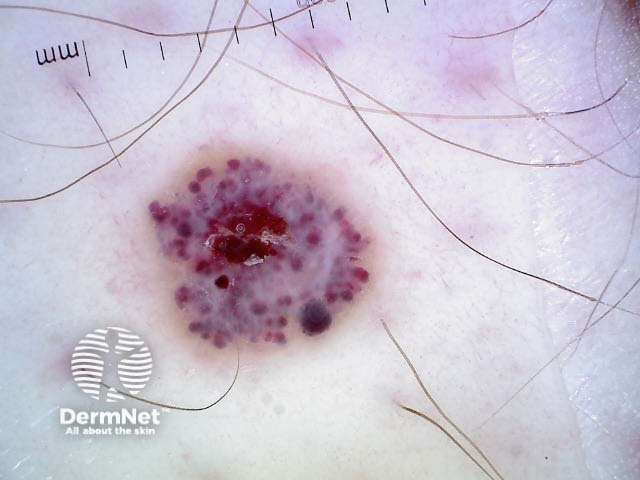
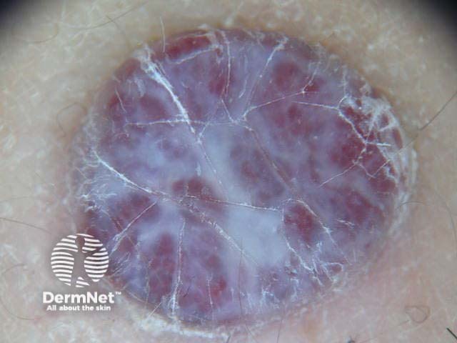
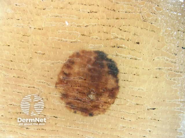
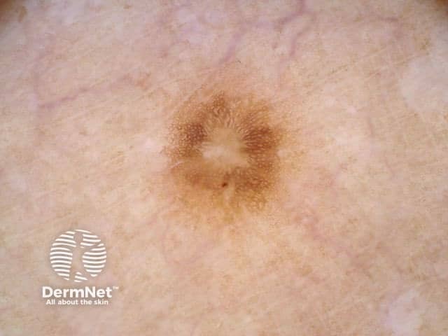
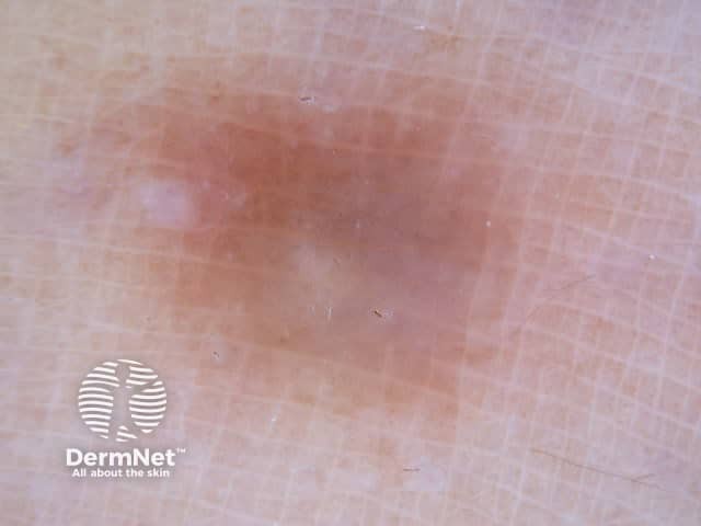
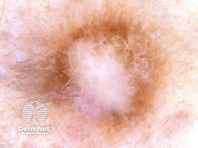
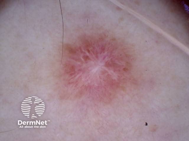
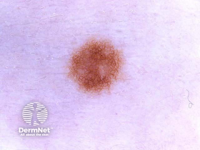
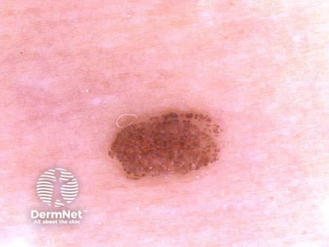
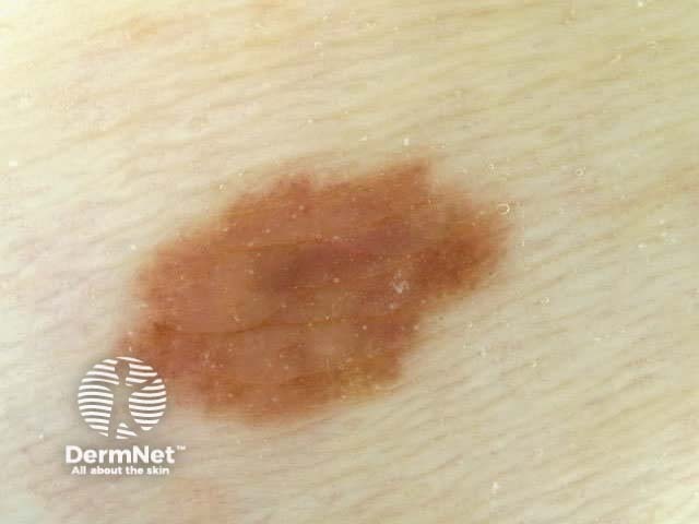
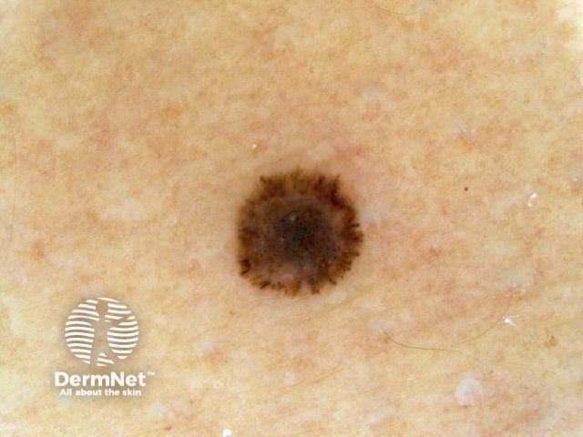
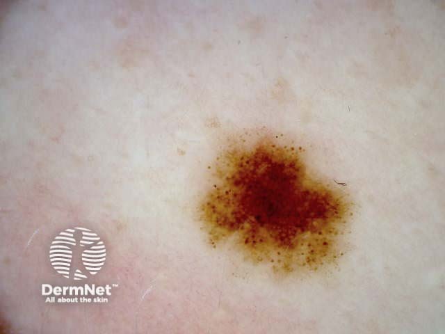
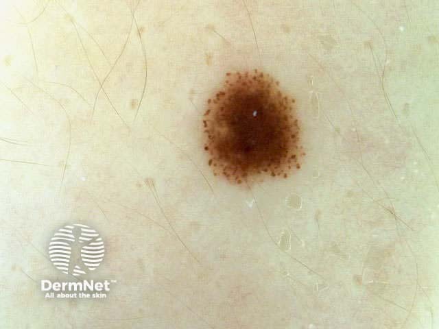
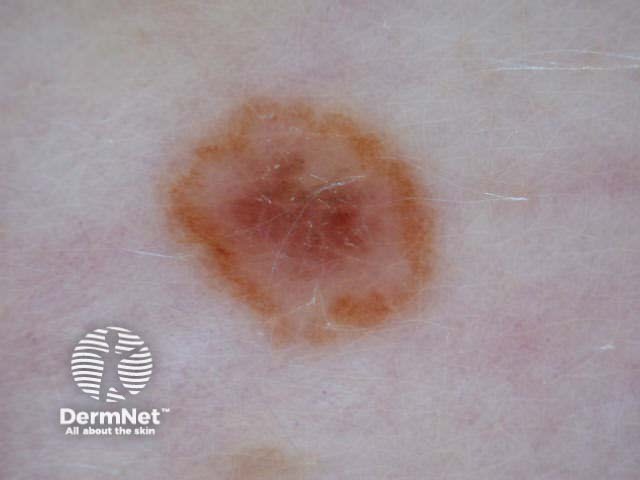
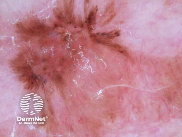
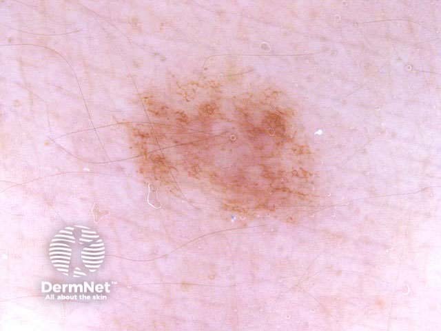
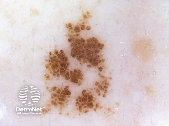
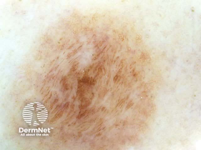
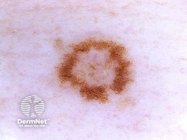
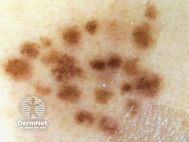
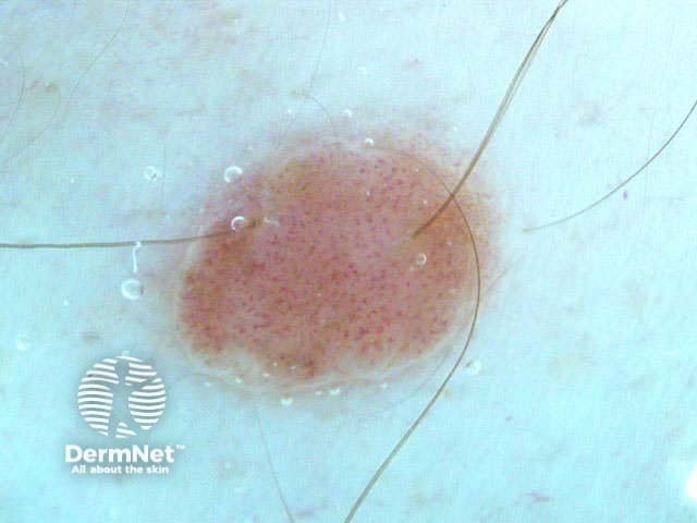
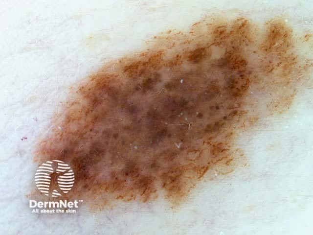
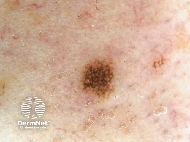
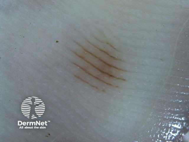
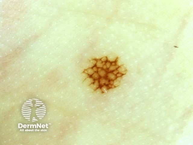
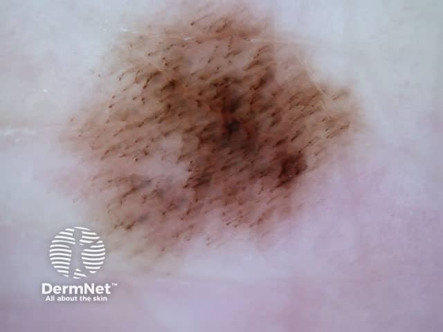
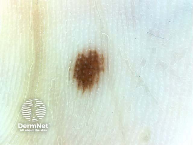
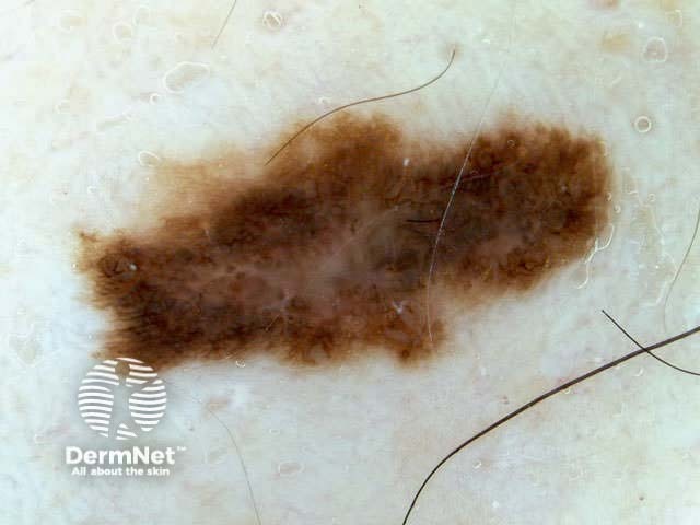
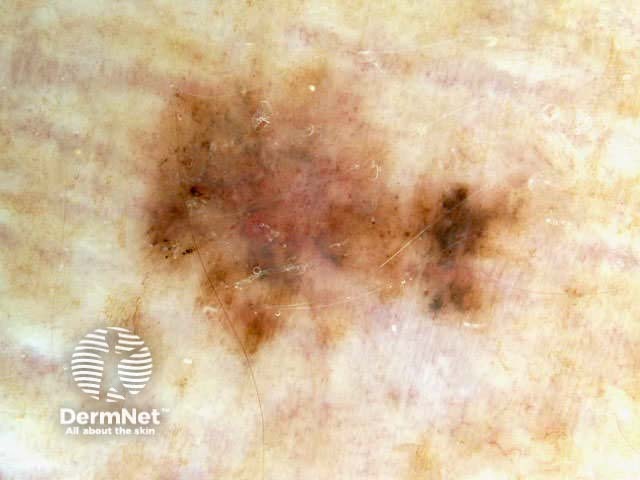
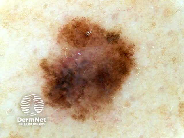
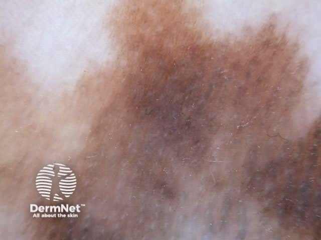
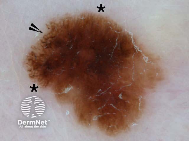
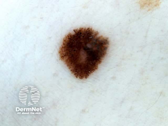
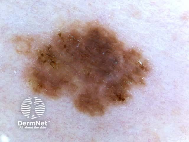
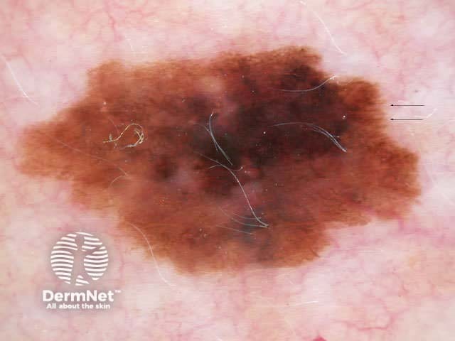
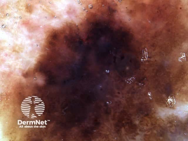
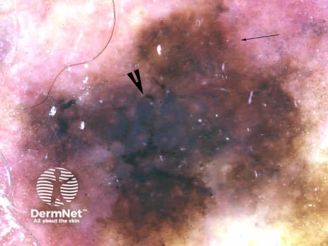
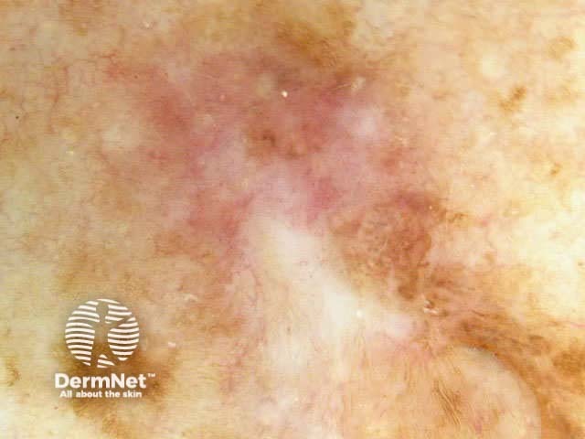
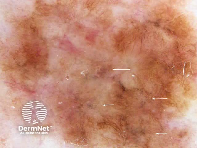
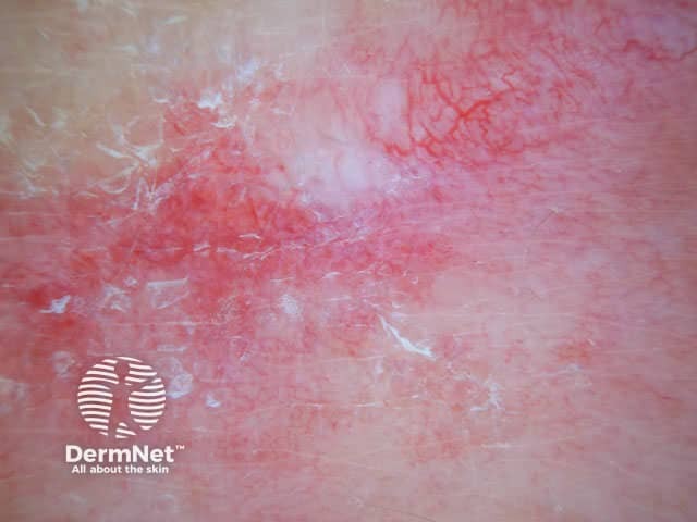
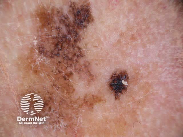
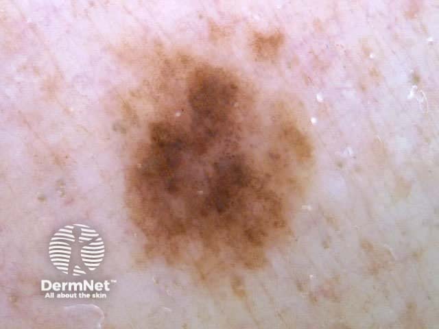
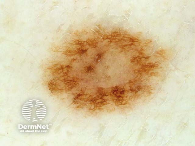
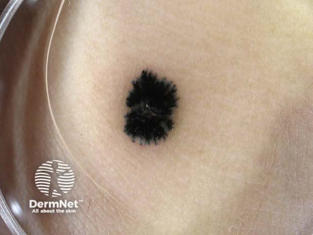
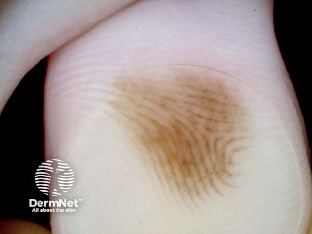
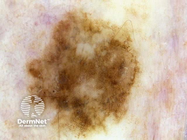
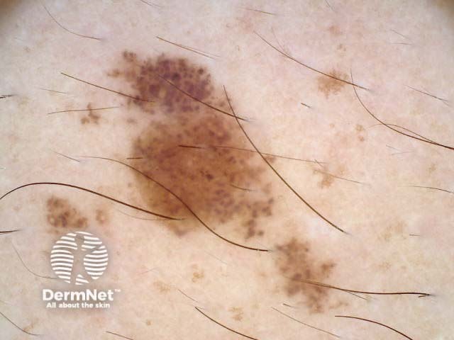
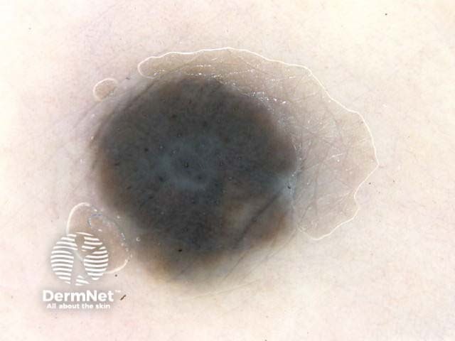
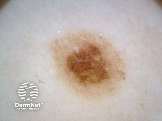
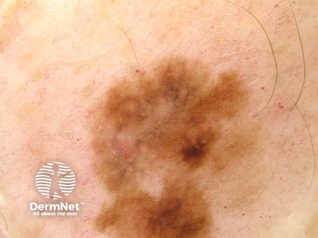
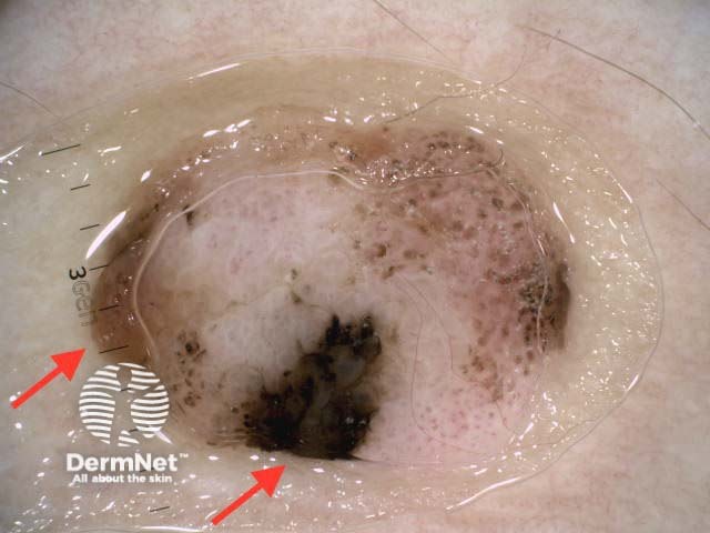
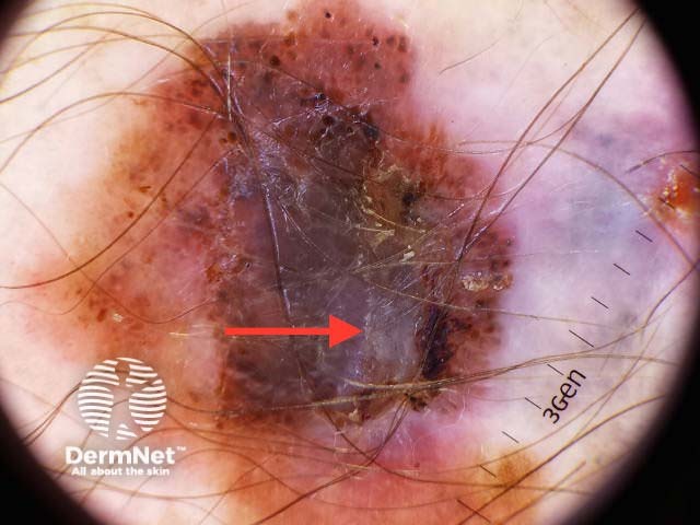
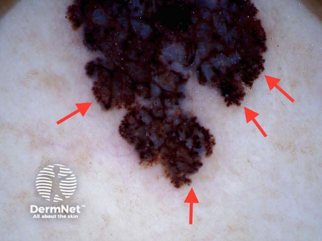
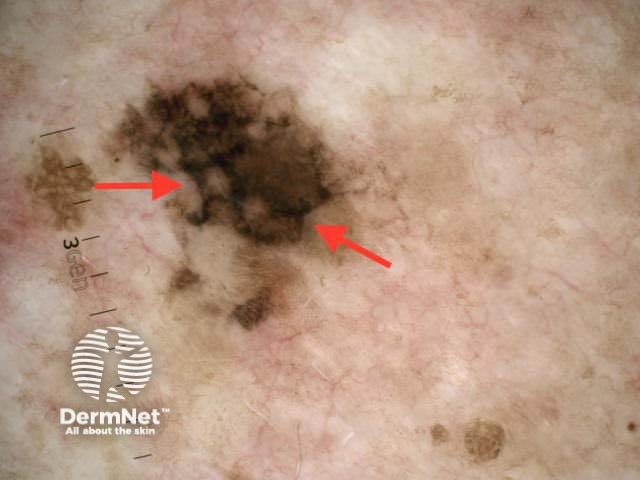
Chaos means the lesion shows more than one pattern, and asymmetry of structure and/or colour on dermatoscopy. This is true for melanoma, basal cell carcinomas and squamous cell carcinomas. Consider excising the lesion if it has one or more dermoscopic clues to malignancy:
All the melanocytic lesions illustrated in the figures below show chaos. Similar chaos and clues can be observed in the basal cell carcinomas illustrated above.

Practice pattern analysis by identifying global and local features in melanocytic lesions. Do the same using modified pattern analysis terminology.
See the DermNet bookstore.

