DermNet provides Google Translate, a free machine translation service. Note that this may not provide an exact translation in all languages

Translate

Dermoscopy

First step algorithm

Created 2008.

Learning objectives

  • Describe the first step algorithm

Introduction

The first step algorithm for dermoscopy distinguishes melanocytic lesions from non-melanocytic lesions. It is used to evaluate pigmented lesions.

Look for specific features of a melanocytic lesion. If these are absent, look for specific features to diagnose pigmented basal cell carcinoma, seborrhoeic keratosis or haemangioma. Also consider whether the lesion could be a viral wart or dermatofibroma (look for the central white patch). If none of these lesions can be diagnosed, treat the lesion as melanocytic (see above).

Melanocytic lesions

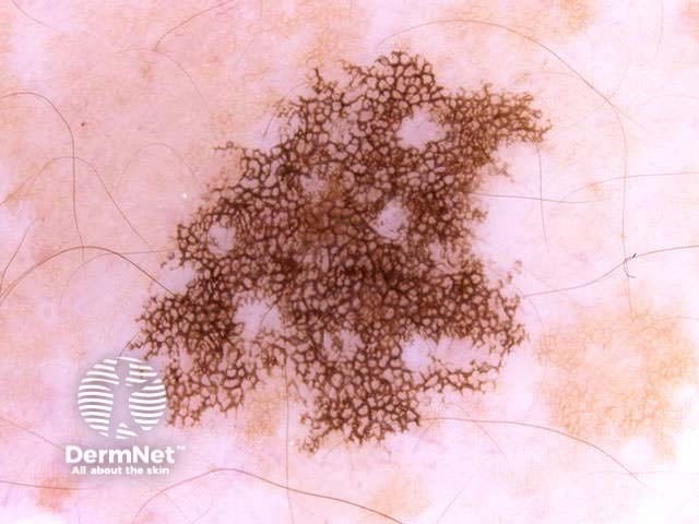
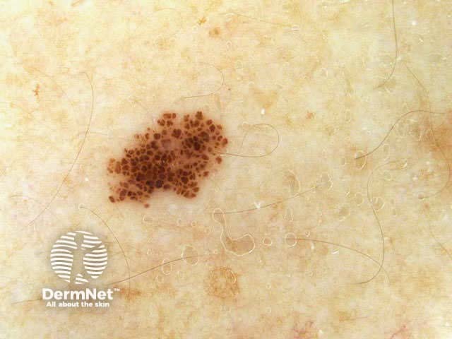
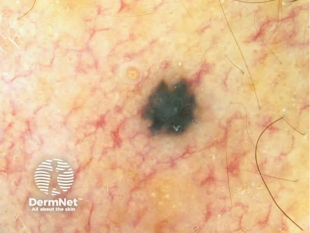
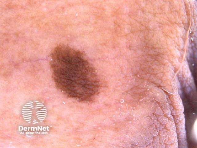
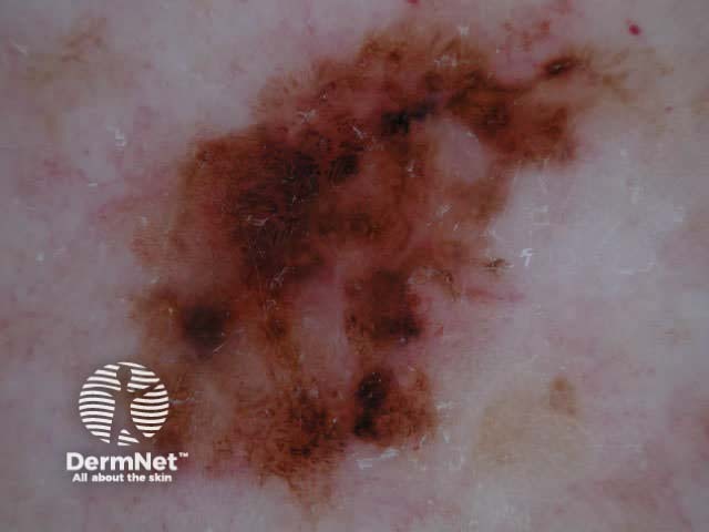
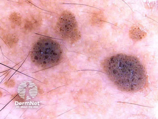
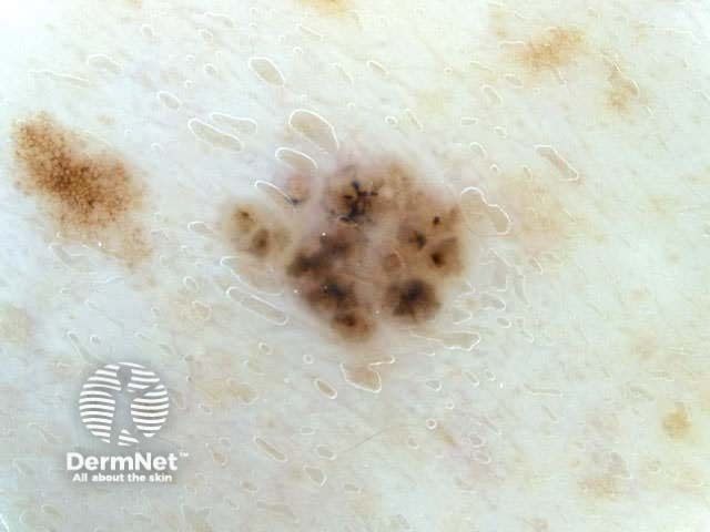
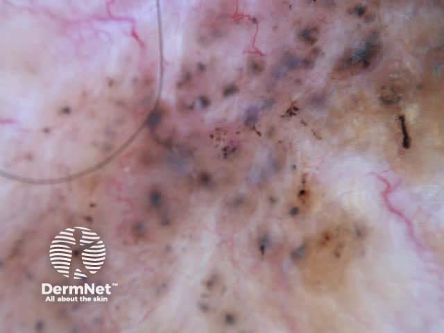
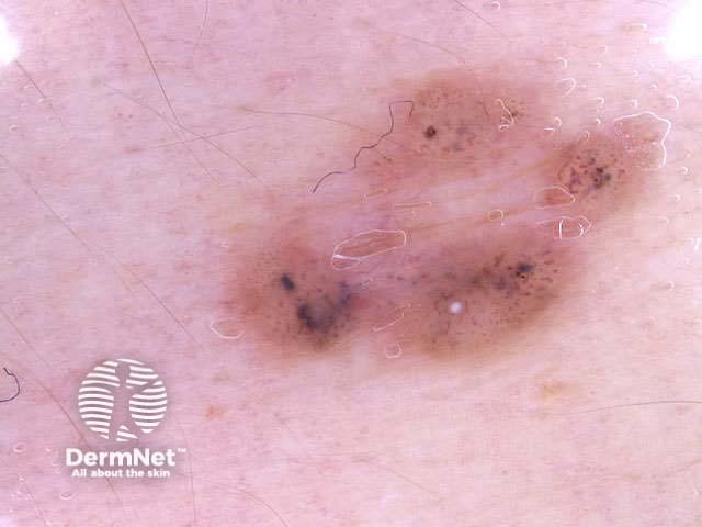
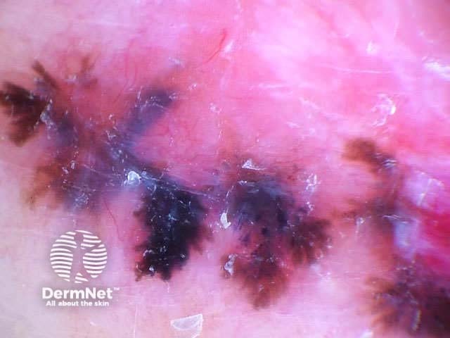
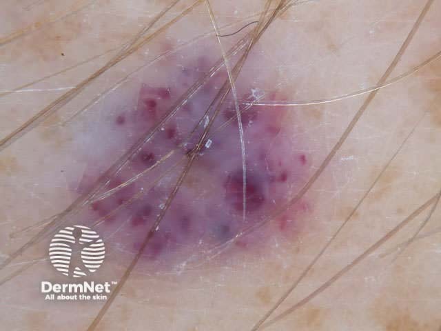
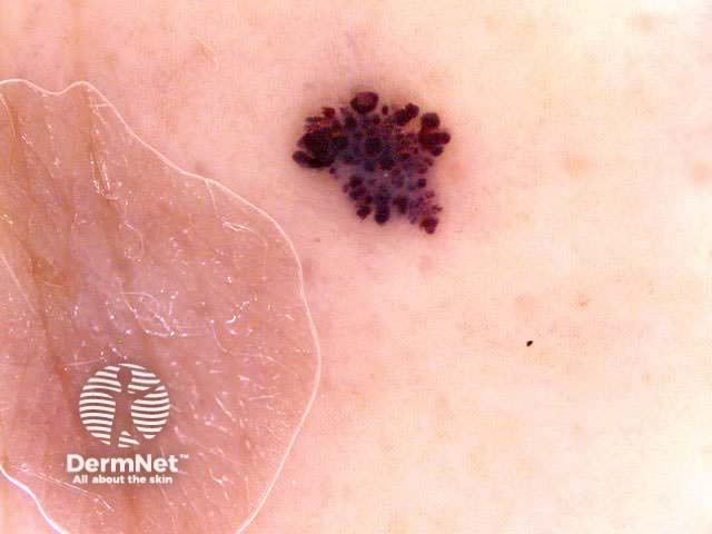
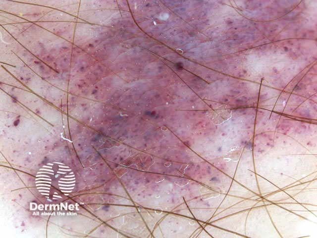
Benign and malignant melanocytic lesions have one or more of the following characteristics:

  • Pigment network or pseudonetwork
    (May also occur in solar lentigo, seborrhoeic keratosis, actinic keratosis, dermatofibroma and accessory nipple)
  • Aggregated globules
  • Streaks
  • Homogeneous blue pigmentation
    (May also occur in haemangiomas, basal cell carcinomas and melanoma metastases)
  • Parallel pattern
  • Or, none of the listed criteria

Non-melanocytic lesions

Seborrhoeic keratoses

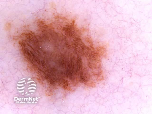
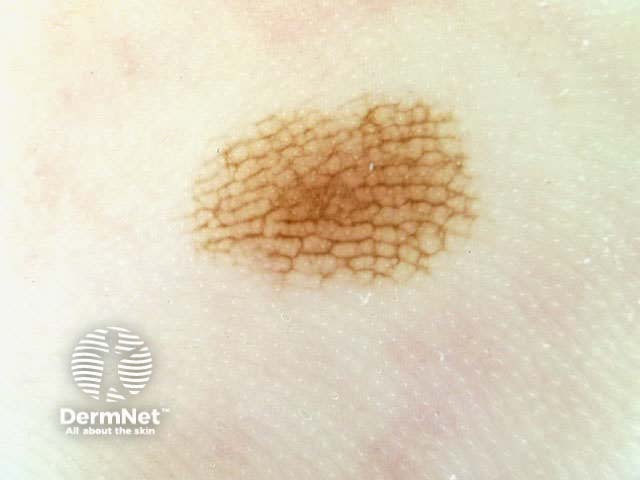
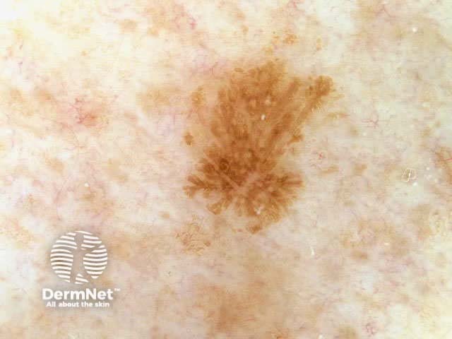
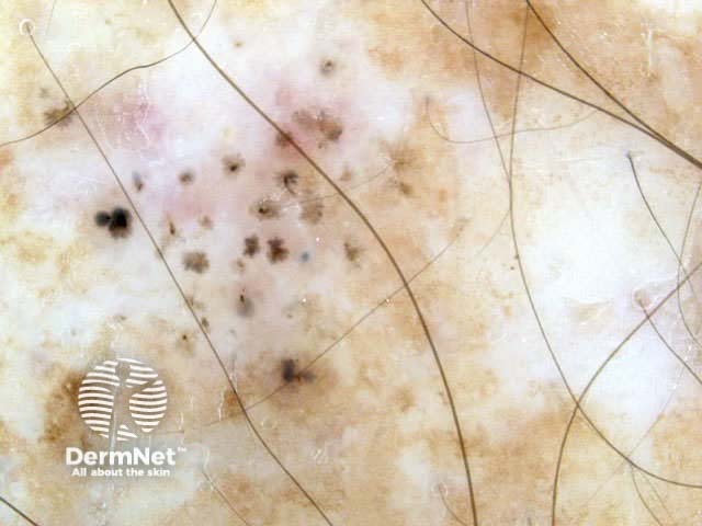
Seborrhoeic keratoses have the following characteristics:

  • Multiple milia-like cysts
  • Comedo-like openings
  • Light-brown fingerprint-like structures
  • Cerebriform pattern

Pigmented basal cell carcinomas

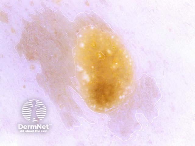
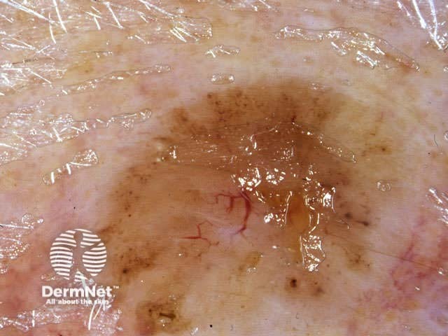
Pigmented basal cell carcinomas have the following characteristics:

  • Absence of pigment network
  • Arborising vessels
  • Leaf-like structures
  • Large blue-grey ovoid nests
  • Multiple blue-grey globules
  • Spoke-wheel areas
  • Ulceration
    (May also occur in melanoma)

Haemangiomas

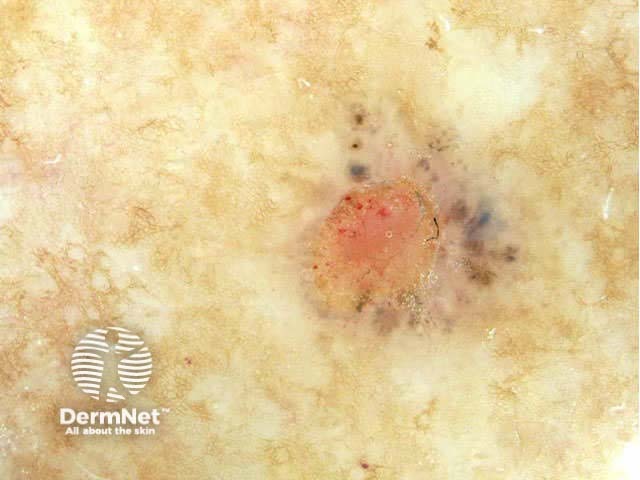
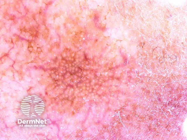
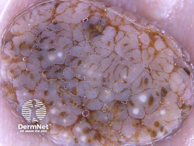
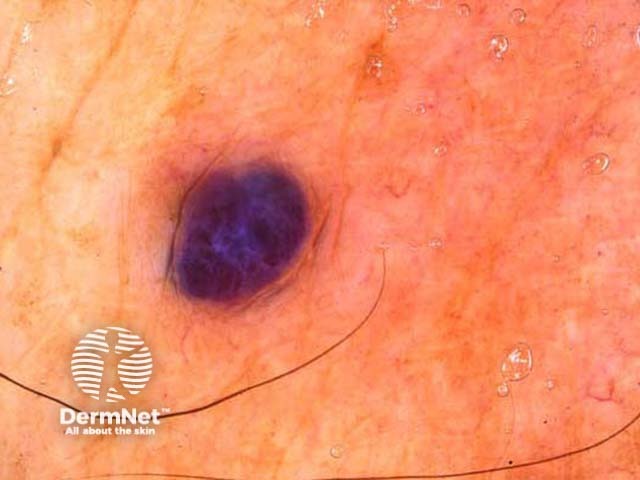
Haemangiomas have the following characteristics:

  • Red-blue lacunes
  • Red-bluish to reddish black homogeneous areas

Second step

The second step is to distinguish benign melanocytic lesions from malignant melanoma using one of the following methods.

  • Pattern analysis
  • ABCD rule
  • Menzies method
  • 7-point rule

If these algorithms appear too complicated, use the 3-point checklist, which is a safe way to identify malignant pigmented lesions.

Activity

Practice identifying melanocytic and non-melanocytic pigmented lesions by dermoscopy.


Related information

References

  • Kittler H. Why the First Step Should Be Abandoned! Arch Dermatol. 2010;146(10):1182-1183. Medline.
  • Dermoscopy of pigmented skin lesions: results of a consensus meeting via the Internet. J Am Acad Dermatol. 2003 May;48(5):679-93. Medline.
  • Braun RP, Rabinovitz HS, Oliviero M, Kopf AW, Saurat JH. Pattern analysis: a two-step procedure for the dermoscopic diagnosis of melanoma. Clin Dermatol. 2002 May-Jun;20(3):236-9. Medline.

On DermNet NZ

Books about skin diseases

See the DermNet NZ bookstore.

Text: Miiskin

Sign up to the newsletter