Introduction Causes Demographics Clinical features Diagnosis Differential diagnoses Treatment Outcome
Discoid eczema is a common type of eczema/dermatitis defined by scattered, well-defined, coin-shaped and coin-sized plaques of eczema. Discoid eczema is also called nummular dermatitis.
Click to see more images of discoid eczema
The cause of discoid eczema is unknown. Some cases are associated with Staphylococcus aureus infection.
The eruption can be precipitated by:
Discoid eczema can affect all age groups. It is slightly more common in older adult males and younger adult females. In males there is an association with chronic alcoholism. Drug-induced discoid eczema can be due to medications that cause skin dryness.
Discoid eczema can occur in association with atopic eczema, eczema craquelé, and secondary eczematisation.
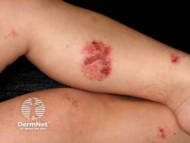
Discoid eczema usually affects the limbs, particularly the legs, but the rash may be widespread. Although often bilateral, the distribution can be asymmetrical especially if related to varicose veins.
There are two clinical forms of discoid eczema:
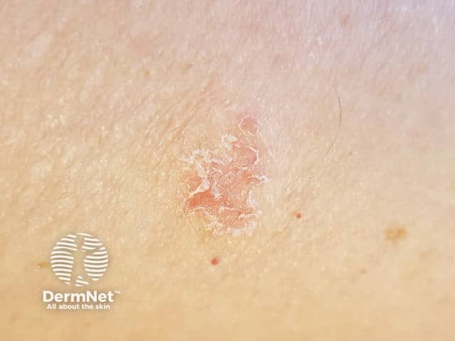
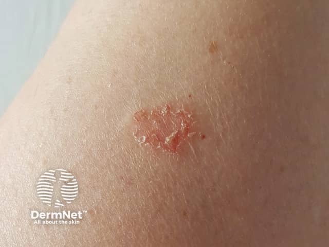
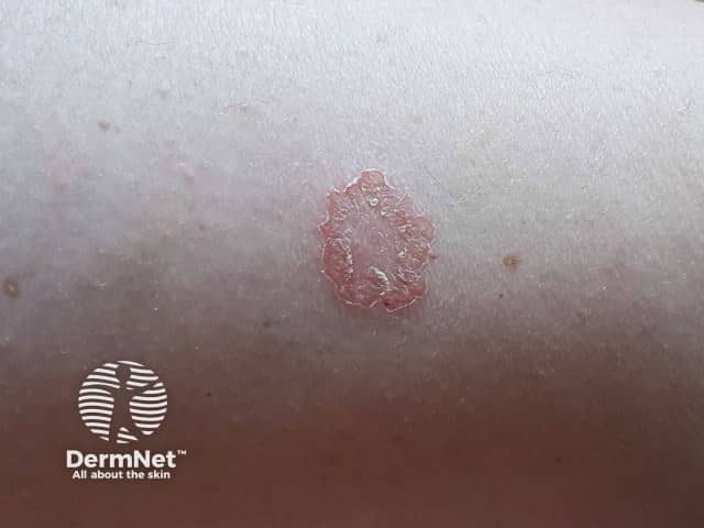
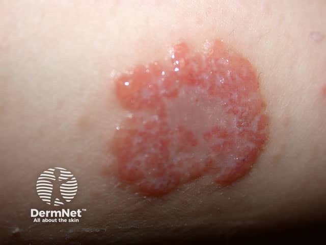
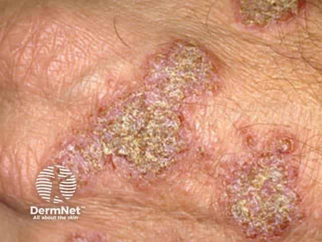
Individual plaques are well circumscribed, mostly 1–3 cm in diameter, and inflamed. The majority of patches are round or oval. The plaques are usually very itchy. The skin between the patches is usually dry and irritable.
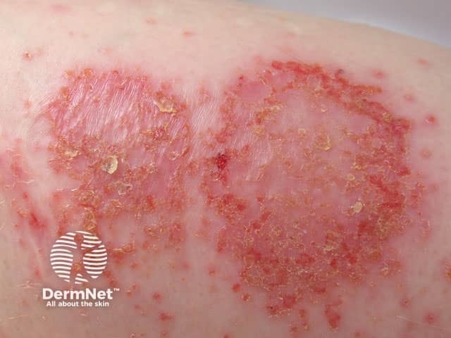
Severe discoid eczema may generalise, with numerous small to large itchy plaques appearing all over the body due to an autoeczematisation reaction.
Patches may clear up without leaving a sign. In dark skin, marks may persist for months as dark brown postinflammatory hyperpigmentation or pale postinflammatory hypopigmentation.
Click to see more images of discoid eczema
In most cases, the appearance of discoid eczema is quite characteristic.
Discoid eczema may resemble other annular skin eruptions including tinea corporis, plaque psoriasis, and pityriasis rosea.
As discoid eczema is associated with loss of skin barrier function, it is important to:
Protect the skin from injury.
This type of dermatitis often starts after minor skin injuries, so careful skin protection is required. If the hands are affected, use gloves and tools to make sure the skin is not irritated by friction, detergents, solvents, other chemicals, or excessive water.
Apply emollients frequently
Emollients include bath oils, soap substitutes and moisturising creams. They can be applied to dermatitis as frequently as required to relieve itching, scaling, and dryness. Emollients should also be used on the unaffected skin to reduce dryness. It may be necessary to try several different products to find one that suits. Many people find one or more of the following helpful: glycerine and cetomacrogol cream, white soft paraffin/liquid paraffin mixed, fatty cream, wool fat lotions, or urea cream.
Avoid allergens
If patch testing has identified contact allergy, exposure to the allergen should be avoided.
Anti-inflammatory treatments include:
Topical steroids
Topical steroids are anti-inflammatory creams or ointments available on prescription to apply just to the patches once or twice daily for 2–4 weeks. They may also be available as steroid impregnated tape. Topical steroids reduce symptoms and clear the dermatitis.
Antibiotics
Antibiotics (eg, erythromycin, flucloxacillin) are often prescribed if the dermatitis is blistered, sticky, or crusted. Sometimes discoid eczema clears completely on oral antibiotics, only to recur when they are discontinued.
Other treatments sometimes prescribed for severe discoid eczema include:
Oral antihistamines
Antihistamine pills may reduce the itch in some patients with discoid eczema. They do not clear the rash.
Ultraviolet radiation (UV) treatment
Phototherapy several times weekly for 6–12 weeks for generalised or widespread discoid eczema can reduce itch and improve the rash.
Steroid injections
Intralesional steroids are sometimes injected into one or two particularly stubborn areas of discoid eczema. This treatment is unsuitable for multiple lesions.
Oral steroids
Systemic steroids are reserved for severe and extensive cases of discoid eczema. They are usually prescribed for a few weeks before continuing topical steroids and emollients on residual dermatitis.
Other oral treatments
Persistent and troublesome discoid eczema is occasionally treated with methotrexate, azathioprine or ciclosporin. These medicines require careful monitoring by a specialist dermatologist. They may be more suitable than long-term systemic steroids.
Discoid eczema tends to be a chronic condition that often relapses especially in cold winter months. Many cases do eventually resolve.

