DermNet provides Google Translate, a free machine translation service. Note that this may not provide an exact translation in all languages

Translate

The complement system

Author: Dr Leeatt Green, Resident Medical Officer, Western Health, Melbourne, Victoria, Australia. DermNet NZ Editor in Chief: Adjunct A/Prof Amanda Oakley, Dermatologist, Hamilton, New Zealand. Copy edited by Gus Mitchell. September 2018.


toc-icon

What is the complement system?

Complement is part of the innate immune system.

Complement incorporates proteins, which circulate in the blood in their inactive form, and some cell membrane receptors [2]. The proteins are mostly synthesised by the hepatocytes in the liver.

The components of complement are systematically named alphanumerically: C1, C2, C3, C3a, C3b, C4, C4a, and so on.

There are three ways to activate the complement system:

  1. The classical pathway, which has two components, globulin (C1) and albumin (C2)
  2. The alternate pathways
  3. Mannose-binding lectin.

All three pathways culminate in the conversion of C3 to C3 convertase [2].  

Skin conditions associated with defects in complement 

The classical complement pathway

When an antibody binds to an antigen, it forms an antigen–antibody immune complex, and this activates the classical complement pathway.

C1 is a protein complex is made up of subunits C1q, C1s, and C1r.

  • Complement is activated when an immune complex binds to C1. Note that only IgM and IgG antibodies can bind to C1.
  • This causes a conformation change in C1, which in turn causes it to activate and cleave C2 into C2a and C2b, and to cleave C4 into C4a and C4b.
  • C2b and C4b then bind together to form a complex called C3 convertase.
  • C3 convertase converts C3 into C3a and C3b.
    • C3a binds to the membranes of mast cells and basophils, which triggers histamine release from the cells.
    • Histamine causes increased vascular permeability and recruits other immune cells to the site of infection.
    • C3b has two functions.
      • It acts as an opsonin to coat cells to alert phagocytes to the cell and makes it easier for these cells to engulf the pathogens
      • It binds to C5 making it easier for C3 convertase to bind to C5.
    • C3 convertase binds to C5; C3b cleaves C5 into C5a and C5b.
    • C5a stimulates histamine release and stimulates chemotaxis.
    • C5b then stimulates the formation of the membrane attack complex.

The complement pathway

The membrane attack complex

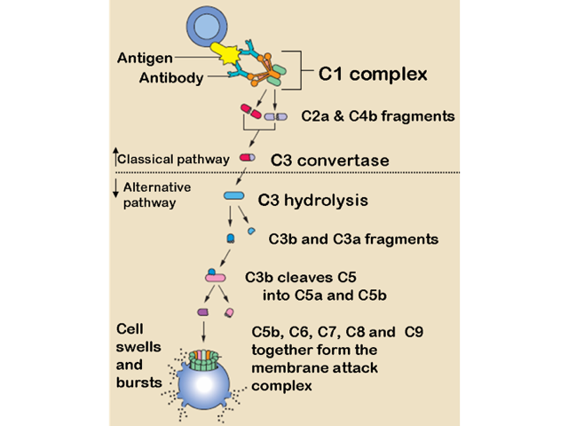
The membrane attack complex is composed of C5b, C6, C7, and C8.

  • This complex binds to a cell surface of a pathogen.
  • It creates a hole in the cell membrane and is surrounded by C9.
  • The cell contents leak out causing the cell to lyse and be destroyed [3].

The alternate pathway

The alternate pathway does not require an antigen–antibody complex.

C3 spontaneously converts to C3a and C3b.

  • In the absence of pathogens, these proteins are degraded [2].
  • If there is a pathogen, C3b binds to a protein in the blood called factor B, forming a C3b–Bb complex.

The C3b–Bb complex does two things.

  1. It is a C3 convertase and activates C3 to form more C3b, thus amplifying the cascade of events.
  2. It can activate C5 to form the membrane attack complex.

Mannose-binding lectin

A lectin is a protein that binds sugars such as mannose.

The mannose-binding lectin pathway involves two proteins:

  1. Mannose-binding lectin (MBL), which doesn’t require antibodies to activate complement
  2. Mannose-binding lectin-associated serine proteinase (MASP).

MBL binds mannose on the surface of bacteria, which causes a conformation change in the structure of MBL that allows MASP to cleave C2 into C2a and C2b, and to cleave C4 into C4a and C4b [4].

C2b and C4b form C3 convertase and amplify the complement system (see alternate pathway).

How is the complement system regulated?

There are regulators to stop the complement system being constantly activated.

  1. C1 inhibitor inhibits C1 and regulates the activity of the classical pathway.
  2. Factor I deactivates C3b.
  3. Factor H inactivates Bb protein and prevents the formation of C3b–Bb complex [4].

Which skin conditions involve defects in the complement system?

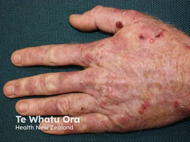
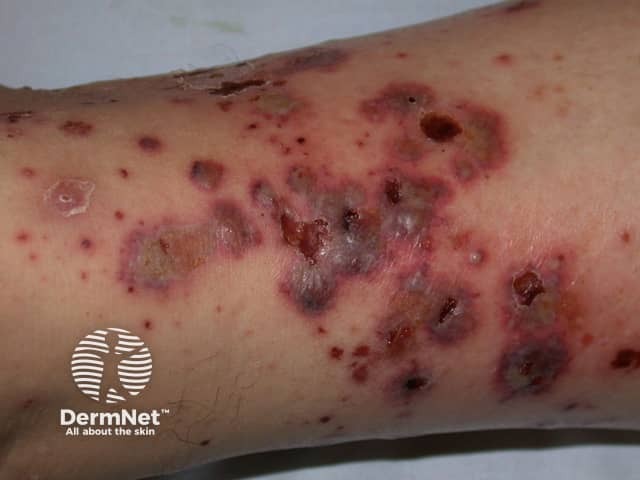
Several skin conditions are associated with defects in the complement system [3].

Blistering skin diseases

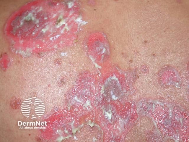
Blistering skin diseases that involve defects in the complement system include:

Vascular diseases

Vascular diseases that involve defects in the complement system include:

Other inflammatory diseases

Other inflammatory diseases that involve defects in the complement system include:

How is complement testing used for diagnosis?

Complement levels can be measured by adding a serum sample into a solution with antigens. Any antibodies bind the antigens to form an immune complex which precipitates out of solution.

The components of the complement system are heat labile, therefore specimens sent for complement assays must be handled carefully. An abnormal result may be due to mishandling during transport of the specimen.


References

  1. Complement system. Wikipedia. 2018. Available at: https://en.wikipedia.org/wiki/Complement_system#cite_note-1 (accessed 20 February 2018).
  2. Gargani Y. Crash course haematology and immunology, 4th edn. London: Mosby, 2015.
  3. Kotnik V. Complement in skin diseases. Acta Dermatovenerol Alp Pannonica Adriat 2011; 20: 3–11. PubMed
  4. Playfair JHL, Chain BM. Immunology at a glance, 10th edn. Hoboken, NJ: Wiley-Blackwell, 2013.

On DermNet

Other websites

Books about skin diseases


Related information

Sign up to the newsletter