Introduction Clinical features Dermoscopic features Histological explanation Differential diagnoses
Achromic naevus, also called naevus depigmentosus or hypochromic naevus, is a pale (hypopigmented) birthmark due to an abnormal clone of melanocytes with an impaired ability to transfer melanosomes to keratinocytes.
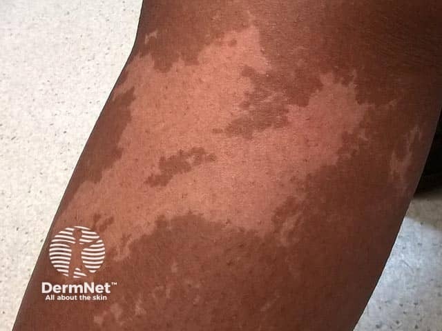
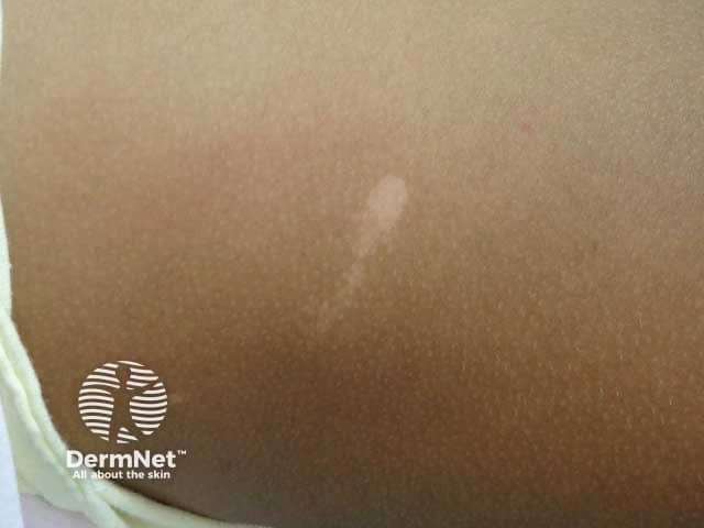
Achromic naevus presents in the neonate or early childhood period as a flat pale hypopigmented macule with a characteristic serrated border.

On dermoscopy, an achromic naevus appears as a white structureless area with an irregular serrated border and pseudopod-like extensions. There is a faint but normal reticular pigment background throughout. Hairs within the lesion are of normal colour with perifollicular pigmentation. Pigmentation in the surrounding skin is normal with no hyperpigmented border.
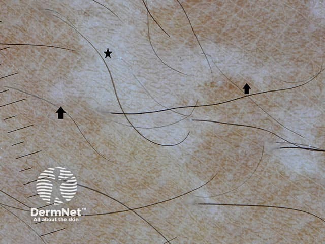
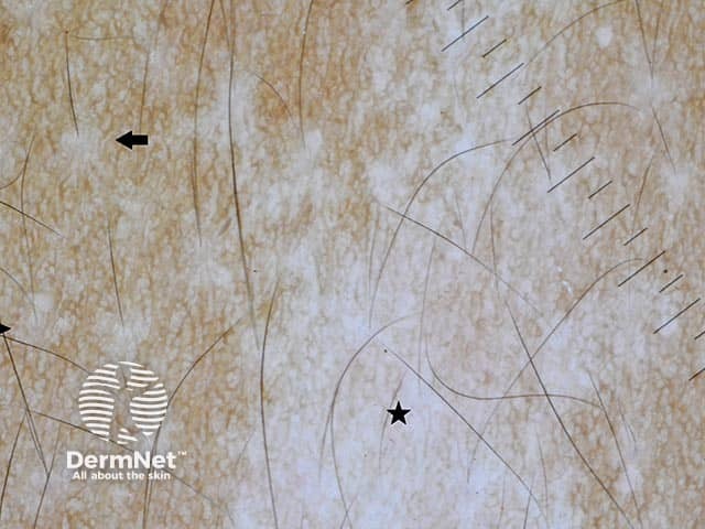
The histology of achromic naevus shows a normal or slightly reduced number of melanocytes with reduced melanosomes in the keratinocytes. The white structureless areas seen on dermoscopy of achromic naevus correspond with reduced melanin in the epidermal keratinocytes. The faint reticular pigment network indicates melanin remaining in the melanocytes.

