

Labial melanotic macule — extra information
Introduction Clinical features Causes Investigations Treatment
What is a labial melanotic macule?
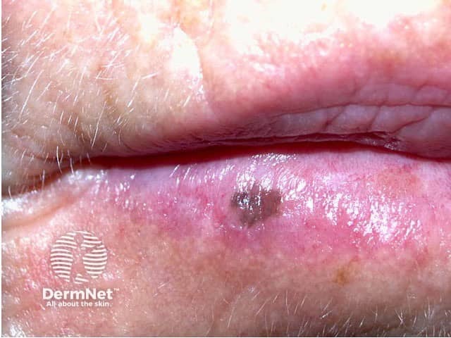
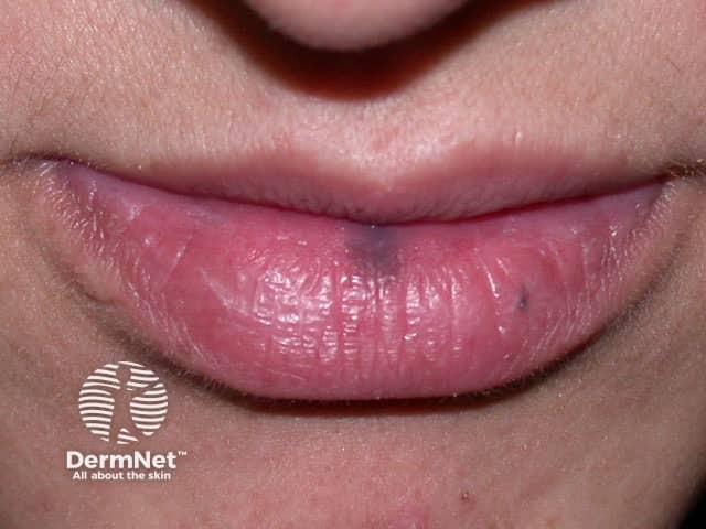
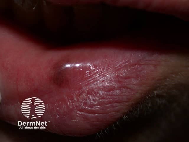
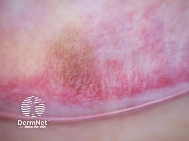
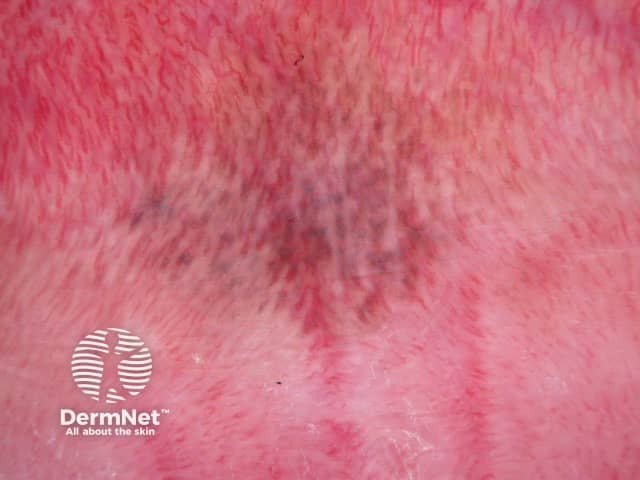
A labial melanotic macule is a well-defined, oval, brown to black, flat patch on the central third of the lower lip. It is the name for a freckle arising on the lip. It is also sometimes called a labial lentigo and when multiple lesions are present, mucosal melanosis.
What are the clinical features of labial melanotic freckle?
Usually solitary, a labial melanotic macule is most commonly seen in adult women but it also occurs in males and in young people. Occasionally the lesion can be on the upper lip.
Size ranges from 1–8 mm. Once developed the lesions usually remain unchanged in size and colour. They can occasionally have an irregular edge and there may be a history of colour change which can cause confusion with other pigmented lesions, including melanoma. Luckily, melanoma is very rare on the lip (but it can occur).
Similar freckles may also occur in areas that are not exposed to the sun:
- Inside the mouth (oral melanotic macules)
- On the vulva in women (vulval labial melanotic macule, vulval melanosis)
- On the penis in men (penile melanotic macule, a penile lentigo).
Labial melanotic macules do not cause any symptoms but their appearance can be a concern to the patient.
What is the cause of labial melanotic macule?
A labial melanotic macule is thought to be provoked by sun exposure, and it is more common in fair-skinned people. However it may also occur in dark-skinned individuals and, as described above, similar lesions can arise in sites that are never sun-exposed. Luckily, melanotic macules are harmless.
What other conditions cause lip pigmentation?
A labial melanotic macule may be confused with another pigmented skin lesion.
- Freckle (ephelides)
- Lentigo simplex
- Solar lentigo
- Venous haemangioma (venous lake)
- Amalgam tattoo
- Junctional melanocytic naevus (a flat mole)
- Lentigo maligna (a form of melanoma in situ)
- Superficial spreading melanoma.
These conditions can be differentiated from labial melanotic macule by a combination of clinical and histological features.
Multiple lesions may be a sign of a widespread skin condition, such as:
- Peutz Jeghers syndrome
- Addison disease
- Laugier-Hunziker syndrome
- Multiple lentiginosis (various syndromes including Noonan syndrome with multiple lentigines).
What investigations should be done?
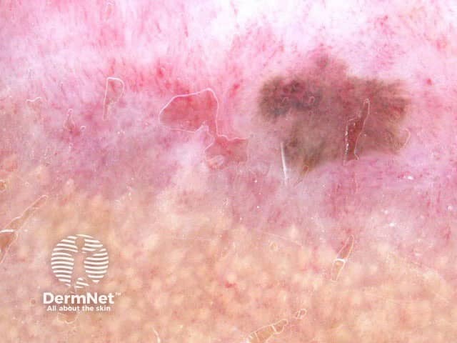
A labial melanotic macule has a characteristic pattern when examined with the magnifying glass or by dermoscopy. Lesions with a typical history and appearance need not be biopsied.
If a skin biopsy is done because the lesion is changing or looks irregular, a labial melanotic macule shows the following features on dermatopathology:
- Increased melanin in the melanocytes and keratinocytes of the basal layer
- Melanophages in the dermal papillae, indicating pigmentary incontinence
- Mild acanthosis without elongation of the rete ridges.
Nuclear atypia is absent and the melanocyte count is normal.
What is the treatment of labial melanotic macule?
Typical lesions can just be observed. Suspicious lesions, including lesions showing progressive change, should be biopsied.
If treatment is requested the macules can be frozen (cryotherapy) or removed using a laser or intense pulsed light. Excision can also be performed but will leave a scar.