Introduction
Classification
Demographics
Causes
Clinical features
Complications
Differential diagnoses
Diagnosis
Treatment
Follow-up
Outcome
Melanoma in situ is an early form of primary melanoma in which the malignant cells are confined to the tissue of origin, the epidermis. It is also known as in-situ melanoma and level 1 melanoma.
As melanoma in situ has no associated mortality, early detection of melanoma in an in-situ phase increases survival from melanoma and leads to less morbidity and decreased costs compared to that associated with more advanced melanoma [1].
Management of melanoma is evolving. For up to date recommendations, refer to Australian Cancer Council Clinical practice guidelines for the diagnosis and management of melanoma.
See more images of melanoma in situ.
Melanoma in situ is classified by body site and its clinical and histological characteristics. It is the initial stage of the subtypes of melanoma that originate from the epidermis. The most common subtypes are:
Rare forms of melanoma that may have an in-situ phase include:
There were 2423 melanoma registrations in New Zealand in 2015. The New Zealand Cancer Registry does not publish the figures for melanoma in situ, but unpublished data suggest that about the same number of people are diagnosed with in-situ melanoma as those diagnosed with invasive melanoma [2].
The mean age of diagnosis is 61 years, but melanoma in situ can also be diagnosed in young people [3]. Mostly it is diagnosed in people who have many melanocytic naevi or in older people with fair skin.
Patients with melanoma in situ may have also been diagnosed with other keratinocytic forms of skin cancer, such as basal cell carcinoma, actinic keratosis, intraepidermal squamous cell carcinoma, and cutaneous squamous cell carcinoma.
Genetic mutations in the DNA of melanocytes are observed in melanoma in situ. These are predominantly due to exposure to ultraviolet radiation.
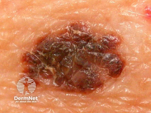
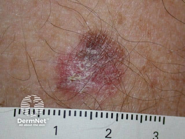
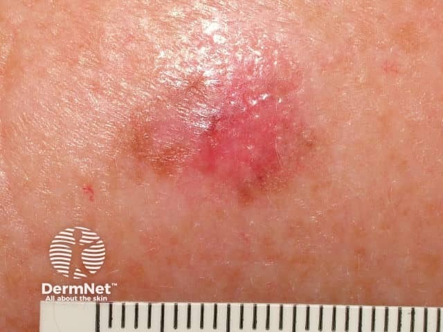
Typically, melanoma in situ is an irregular pigmented patch of skin. It often has the ABCDE criteria:
The body site and other clinical features of melanoma in situ depend on the subtype of melanoma (see above). In general terms, melanoma in situ is macular (flat). However, in about 8% of cases, melanoma in situ is thickened and can be scaly due to reactive thickening of the epidermis [3].
Untreated, melanoma in situ slowly enlarges. Some in-situ melanomas develop foci (a centre of a morbid process) or a more potentially dangerous, invasive form of melanoma.
Differential diagnoses for melanoma in situ include invasive melanoma, other forms of skin cancer, and benign skin lesions, such as a melanocytic naevus or lentigo (these may have been clinically described as atypical naevus or atypical solar lentigo).
Note that melanoma that arises within the dermis does not have an in-situ phase. Dermal subtypes of melanoma include:
Melanoma in situ may be suspected clinically or by dermoscopy.
Diagnosis is confirmed by histological examination of the tumour and finding malignant melanocytes confined to the epidermis and epidermal adnexal structures. Breslow thickness is not reported for melanoma in situ.
Multiple sections through the specimen should be examined to ensure there are no areas of invasive disease. Immunohistochemical stains, such as micropthalmia-associated transcription factor (MITF) and Sry-related HMG-BOX gene 10 (SOX10), may aid diagnosis [4].
Melanoma in situ is treated by excision biopsy. A special tissue-sparing technique may be used for a large melanoma in situ, such as Mohs micrographic surgery or staged mapped excisions [2].
When surgical margins are narrow, a second surgical procedure is undertaken, including a 5–10mm clinical margin of normal skin, to ensure complete removal of the melanoma. This is known as wide local excision.
Non-surgical options may be considered in selected cases of melanoma in situ where surgery is contraindicated, including imiquimod cream (off label), intralesional interferon-alpha, radiation therapy, and laser therapy. Recurrence rates are high with these second-line treatments.
Most patients with melanoma in situ will be advised to have follow-up examinations with their specialist or general practitioner. The main focus will be a total body skin examination, because patients with a melanoma in situ have eight times the risk of developing another in-situ or invasive primary melanoma compared to matched individuals without melanoma in situ.
Patients with melanoma in situ have the same life expectancy as the general population. Further problems are rare from melanoma in situ because the malignant cells within the epidermis have no metastatic potential. However, a small focus of invasive disease may have beeen missed due to the impracticability of evaluating every part of a large skin lesion.
Melanoma in situ occasionally recurs at the same site, requiring further surgery.
Management of melanoma is evolving. For up to date recommendations, refer to Australian Cancer Council Clinical practice guidelines for the diagnosis and management of melanoma.

