DermNet provides Google Translate, a free machine translation service. Note that this may not provide an exact translation in all languages

Translate

Laser therapy in skin of colour

Author: Dr Ramez Barsoum, Resident Medical Officer, Princess Alexandra Hospital, Brisbane, QLD, Australia. DermNet NZ Editor in Chief: Adjunct A/Prof Amanda Oakley, Dermatologist, Hamilton, New Zealand. Copy edited by Gus Mitchell/Maria McGivern. June 2019.


toc-icon
Table of contents
arrow-right-small

What is skin of colour?

Skin of colour refers to non-white skin types, with a particular emphasis on Fitzpatrick skin phototypes V and VI. It is characterised by increased epidermal melanin (a brown pigment), more widely distributed melanosomes (the melanin-containing granules within melanocytes), changing melanocyte response, and overactive fibroblasts.

Skin of colour

How does skin of colour differ from white skin?

Since melanin absorbs and scatters the energy transmitted from ultraviolet radiation (UVR), persons with skin of colour experience less epidermal damage after exposure to UVR and show fewer signs of photoageing than people with lighter skin types [1,2].

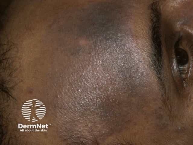
Skin of colour almost always develops pigmentary changes when exposed to injury or inflammation (postinflammatory hyperpigmentation and postinflammatory hypopigmentation), whereas pigmentary changes are uncommon in white skin [3].

Melanin can act as a competitive chromophore (a coloured molecule that absorbs transmitted energy), which increases the risk of side effects after epidermal injury by a laser [4].

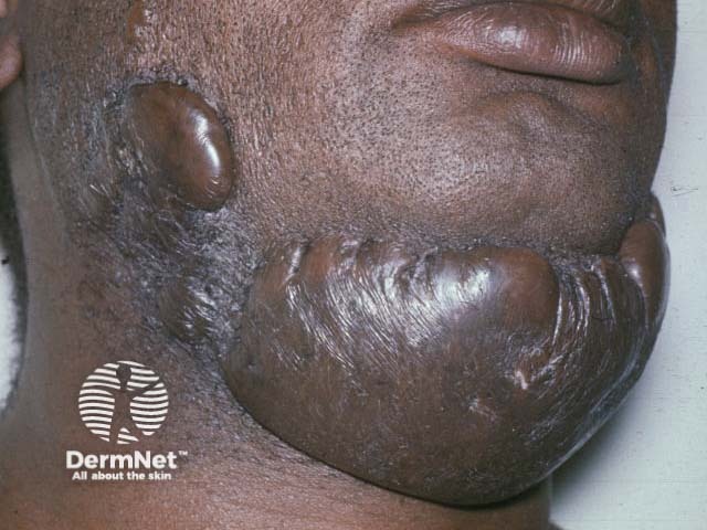
People with skin of colour have a higher prevalence of hypertrophic scarring and keloids after injury than those with white skin due to genetic factors associated with hyperactive fibroblasts [2,3].

What are the indications for laser therapy in skin of colour?

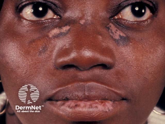
The medical indications for laser therapy are similar whatever the colour of the skin. However, women of colour often seek treatment for hyperpigmentation and uneven skin colour as these are common aesthetic concerns [2].

Laser therapy may also be used to remove dark, coarse, terminal hairs, which may lead to pseudofolliculitis barbaefolliculitis barbae, folliculitis keloidalis nuchae, and folliculitis decalvans — conditions that are more common in skin of colour [5].

Laser hair removal in skin of colour

The safest laser for hair removal in individuals with skin of colour is the long-pulsed neodymium-doped yttrium aluminium garnet (Nd:YAG) laser.

  • Its wavelength (1064 nm) is at the end of the absorption spectrum of melanin.
  • Nd:YAG laser results in sufficient thermal injury to dark coarse hairs and spares the epidermis [6].
  • It allows energy to be delivered slowly, resulting in heat dissipation and cooling, and minimising damage to the epidermis.

The use of the alexandrite laser (wavelength of 755 nm) has not been extensively studied in skin of colour. It has been reported to cause blistering in patients with Fitzpatrick skin types V and VI [7].

Use of a diode laser (wavelength 800 nm) has been reported to be mainly safe with low rates of complications, including the occurrence of transient blistering and pigment alteration [8].

Resurfacing acne scars

Acne scars may be treated with an ablative carbon dioxide laser or erbium-doped yttrium aluminium garnet (Er:YAG) laser, but these are best avoided in skin types V and VI as thermal injury commonly causes postinflammatory hyperpigmentation [9].

Non-ablative Nd:YAG laser has fewer side effects compared to ablative laser resurfacing and produces comparable results.

Tattoo removal

Few studies on the removal of tattoos in skin of colour have been reported. Charcoal-based blue/black religious tattoos in Ethiopian patients with skin types V and VI have been removed using a Q-switched Nd:YAG [10]. In this study, almost half of the patients developed mild postinflammatory hyperpigmentation lasting between 2 and 4 months.

Removal of tattoos in skin of colour can be difficult and unpredictable because of the epidermal melanin, which absorbs the transmitted energy and prevents it from reaching the ink in the dermis.

What can help improve the outcomes of laser therapy in skin of colour?

Cooling

Cooling is used to protect the epidermis from thermal burn [11].

  • Contact cooling, which relies on conduction, can be active (eg, through the use of sapphire laser windows or copper tips) or passive (eg, through the use of ice cubes).
  • Non-contact cooling uses cold air convection or cryo-cooling.

Technique

Epidermal injury should be monitored carefully in skin of colour.

  • A Nd:YAG laser with a long wavelength and wider pulse times is often the most suitable device for use in skin of colour.
  • A test pulse or pulses can be used to check the immediate effects of the laser on the skin.
  • Multiple, short sessions can also help reduce epidermal damage compared to single or longer sessions [12].

Education

Patient expectations should be realistic, and they should be informed about the risk of complications and side effects associated with laser therapies [2].


References

  1. Montagna W, Carlisle K. The architecture of black and white facial skin. J Am Acad Dermatol 1991; 24: 929–37. DOI: 10.1016/0190-9622(91)70148-u. PubMed
  2. Grimes PE. Skin and hair cosmetic issues in women of color. Dermatol Clin 2000; 18: 659–65. DOI: 10.1016/s0733-8635(05)70217-5. PubMed
  3. Szabo G, Gerald AB, Patnak MA, Fitzpatrick TB. Racial differences in the fate of melanosomes in human epidermis. Nature 1969; 222: 1081–2. DOI: 10.1038/2221081a0. PubMed
  4. Kushikata N, Negishi K, Tezuka Y, Takeuchi K, Wakamatsu S. Non-ablative skin tightening with radiofrequency in Asian skin. Lasers Surg Med 2005; 36: 92–7. DOI: 10.1002/ism.20136. PubMed
  5. Ross EV, Cooke LM, Timko AL, Overstreet KA, Graham BS, Barnette DJ. Treatment of pseudofolliculitis barbae in skin types IV, V, and VI with a long-pulsed neodymium: yttrium aluminum garnet laser. J Am Acad Dermatol 2002; 47: 263–7. DOI: 10.1067/mjd.2002.124081. PubMed
  6. Alster TS, Bryan H, Williams CM. Long-pulsed Nd:YAG laser-assisted hair removal in pigmented skin: a clinical and histological evaluation. Arch Dermatol 2001; 137: 885–9. PubMed
  7. Weisberg NK, Greenbaum SS. Pigmentary changes after alexandrite laser hair removal. Dermatol Surg 2003; 29: 415–9. DOI: 10.1046/j.1524-4725.2003.29098.x. PubMed
  8. Shah GK. Efficacy of diode laser for treating acne keloidalis nuchae. Indian J Dermatol Venereol Leprol 2005; 71: 31–4. DOI: 10.4103/0378-6323.13783. PubMed
  9. Fisher GH, Geronemus RG. Short-term side effects of fractional photothermolysis. Dermatol Surg 2005; 31: 1245–9. DOI: 10.1111/j.1524-4725.2005.31934. PubMed
  10. Grevelink JM, Duke D, van Leeuwen RL, Gonzalez E, DeCoste SD, Anderson RR. Laser treatment of tattoos in darkly pigmented patients: efficacy and side effects. J Am Acad Dermatol 1996; 34: 653–6. DOI: 10.1016/s0190-9622(96)80068-5. PubMed
  11. Chiu CH, Chan HH, Ho WS, Yeung CK, Nelson JS. Prospective study of pulsed dye laser in conjunction with cryogen spray cooling for treatment of port wine stains in Chinese patients. Dermatol Surg 2003; 29: 909–15. DOI: 10.1046/j.1524-4725.2003.29255.x. PubMed
  12. Adrian RM, Shay KP. 800 nanometer diode laser hair removal in African American patients: a clinical and histologic study. J Cutan Laser Ther 2000; 2: 183–90. DOI: 10.1080/146288300750163754. PubMed

On DermNet

Books about skin diseases


Related information

Sign up to the newsletter