Lipomas are common, usually asymptomatic tumours composed of mature adipose tissue. They have a predilection for the subcutaneous tissue of the trunk and extremities but have been described in a wide range of anatomic locations. They occur in multiplicity in several distinct clinical syndromes.
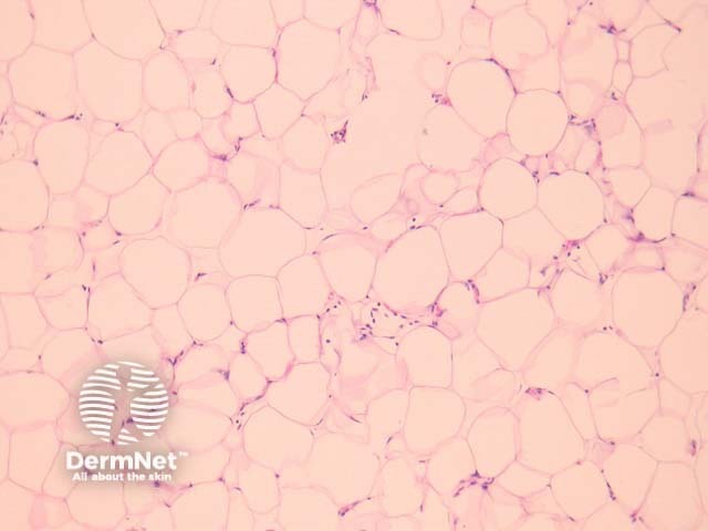
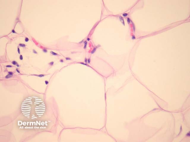
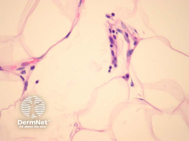
Sections show mature adipose tissue (figures 1-3). The fat contains few small capillaries within thin fibrous strands. A thin fibrous capsule is often seen.
Fat necrosis and other inflammatory changes may be seen when lipomas are traumatised.
Cytogenetic abnormalities can be identified in approximately half of lipomas but are generally not required for diagnostic purposes. Characteristic rearrangements involve chromosomes 12, 6, and 13.
Normal fat — Distinction from normal fat can sometimes be difficult. Circumscription and encapsulation of lobules favours a lipoma. Clinical history may be essential, especially when dealing with emulsified fat specimens (liposuction specimens from lipomas or weight loss procedures).
Well-differentiated liposarcoma — Some areas of well-differentiated liposarcoma can be exceedingly bland and mimic a lipoma. Liberal sampling and histologic examination of fatty tumours is recommended to search for atypical areas.

