Introduction
Histology
Special studies
Differential diagnoses
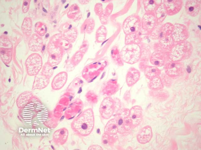
The hibernoma is derived from brown fat. They occur in the subcutaneous or deeper soft tissues.
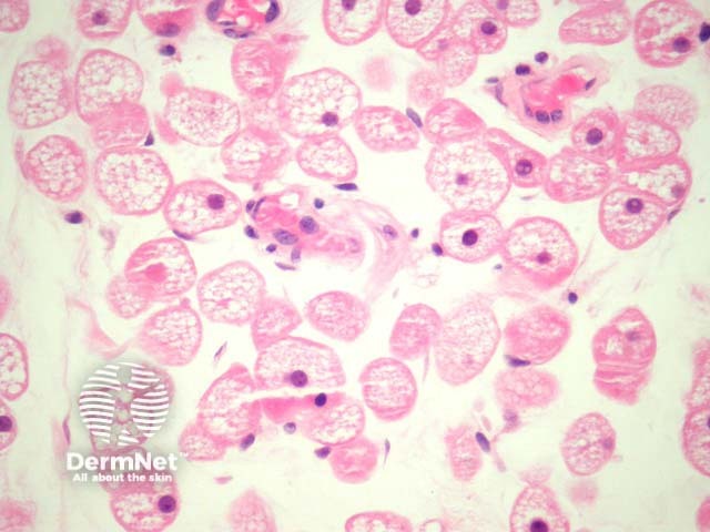
Sections show a highly characteristic lesion composed of cells with a markedly enlarged cytoplasm filled with course vacuoles and eosinophilic granules (figures 1,2).
None are generally needed. Cytogenetic studies of hibernoma have shown deletions on chromosomes 10 and 11 but are not required for diagnosis.
Normal brown fat — Brown fat may be seen incidentally in paediatric soft tissues. Physiologic brown fat does not form a discrete mass.
Granular cell tumour, rhabdomyomas — These tumours contain eosinophilic granules in the cytoplasm. Intracytoplasmic vacuoles are not seen in these tumours.

