Main menu
Common skin conditions

NEWS
Join DermNet PRO
Read more
Quick links
Author: Dr Lauren Thomas, RMO, Royal Darwin Hospital, Darwin, NT, Australia. DermNet Editor in Chief: Adjunct A/Prof. Amanda Oakley, Dermatologist, Hamilton, New Zealand. Copy edited by Gus Mitchell. October 2019.
Introduction Demographics Causes Clinical features Diagnosis Treatment Outcome
Multicentric reticulohistiocytosis is a very rare multisystem arthropathic form of reticulocytosis. Reticulohistiocytoses are a type of non-Langerhans cell histiocytosis. Multicentric reticulohistiocytosis is characterised by skin and mucosal lesions, and arthritis [1].
Multicentric reticulohistiocytosis is referred to as lipoid dermatoarthritis, lipoid rheumatism, and giant cell reticulohistiocytosis.
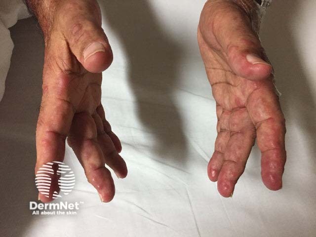
Multicentric histiocytosis
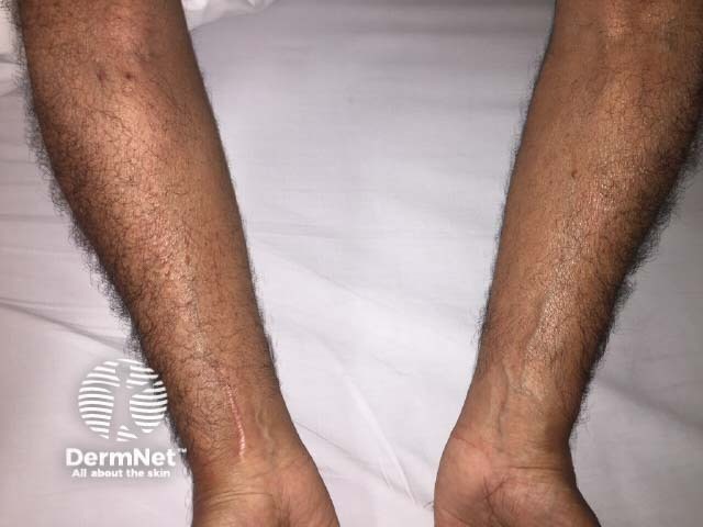
Multicentric histiocytosis
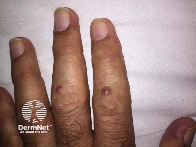
Multicentric histiocytosis
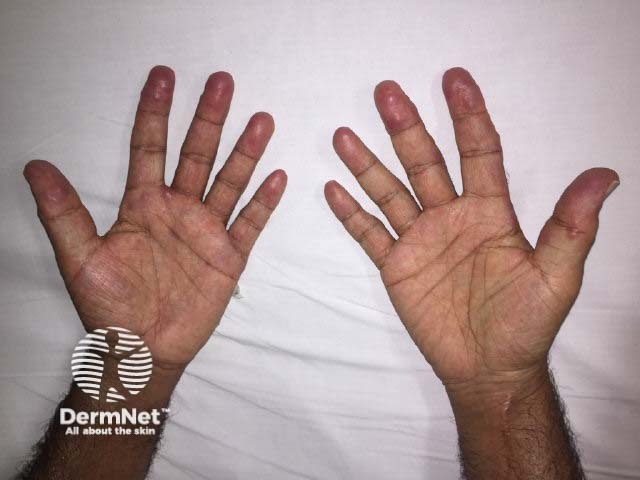
Multicentric histiocytosis
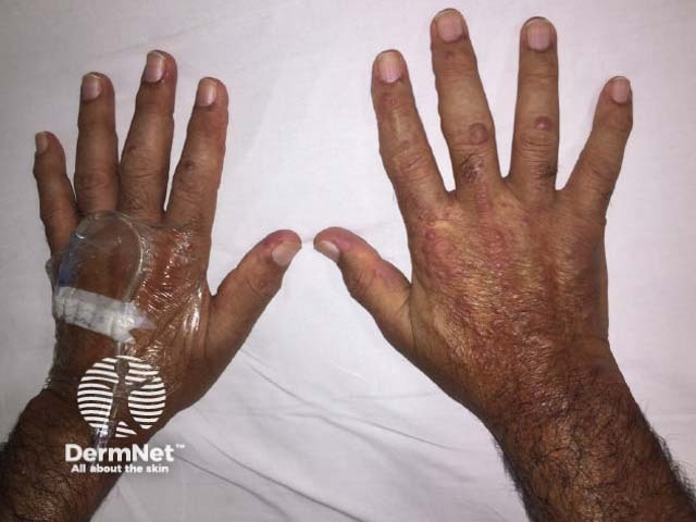
Multicentric histiocytosis
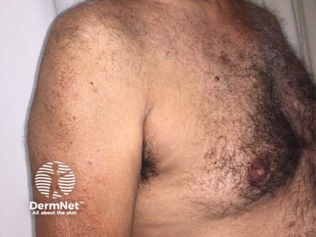
Multicentric histiocytosis
Multicentric reticulohistiocytosis is more common in women than men, with a ratio of 3:1 [1]. The mean age of people affected by multicentric reticulohistiocytosis is 47 years (age range 8-74 years) [5].
In approximately 1 in 4 cases, multicentric reticulohistiocytosis is associated with an internal malignancy [1]. The most common malignancies associated with multicentric reticulohistiocytosis are carcinomas of the lung, stomach, breast, cervix, colon, and ovary.
The cause of multicentric reticulohistiocytosis is unknown. Molecular abnormalities have been reported in the MAP2K1, TT2 and other pathways [5]. Whether multicentric reticulohistiocytosis is a true paraneoplastic disorder is controversial.
Multicentric reticulohistiocytosis is also associated with:
Multicentric reticulohistiocytosis results in papules, nodules, and severe and rapidly destructive arthritis. It can involve the bones, tendons, muscles, joints, and solitary organs (eg, thyroid, eye, lung, kidney, and liver) [1].
Arthritis most commonly involves the fingers, hands, knees, and shoulders, but any joint can be affected. This is a symmetrical progressive arthritis that can be associated with joint stiffness and swelling. The symptoms of arthritis may wax and wane, but can rapidly become severe and lead to joint destruction and deformity in about 45% of patients. Importantly, this can be asymptomatic at the time of presentation, and hence needs to be looked for on radiology.
Multicentric reticulohistiocytosis skin lesions most commonly occur on the upper half of the body, especially the face, ears, mucosal surfaces (lips, tongue, gums, nostrils, throat, eyelids), hands, and forearms.
Dermatoscopy of multiple papules in one patient has been reported. All showed the 'setting-sun sign' of a yellowish hue centrally and a pink-orange rim. Most also had brown reticular structures and less commonly a central white scar-like change were seen [6].
Multicentric reticulohistiocytosis can be accompanied by systemic symptoms, including:
Reticulocytosis is diagnosed from the characteristic histology of a skin biopsy, showing:
The specific diagnosis of multicentric reticulohistiocytosis is made when radiology shows the typical erosive arthritis especially in the hands.
Histiocytes and multinucleated giant cells can also be found in joints and occasionally in internal organs.
Blood tests are not diagnostic; they may include routine haematological investigations, lipid profile, thyroid profile, C-reactive protein (CRP), and rheumatoid factor. Most patients with multicentric reticulohistiocytosis have mild anaemia; 30–60% of patients have hyperlipidaemia and an elevated erythrocyte sedimentation rate (ESR)/C-reactive protein (CRP) [2,3].
A positive tuberculin skin test is found in 12–50% of patients with multicentric reticulohistiocytosis and is more common where tuberculosis is endemic [3].
Once the diagnosis of multicentric reticulohistiocytosis has been made, a thorough history and physical examination should be undertaken with age-appropriate screening tests for malignancy, including a chest x-ray and ultrasound scan of the abdomen/pelvis [1].
There is no specific or curative treatment for multicentric reticulohistiocytosis. The following have been reported to be helpful:
In many patients, multicentric reticulocytosis can go into remission after an average course of 8 years; however, by this time considerable joint destruction may have occurred. Arthritis mutilans may develop in 50% of cases [2]. Patients are left with crippling, deformed joints and a disfigured facial appearance.