

Naevus lipomatosus superficialis — extra information
Introduction Demographics Causes Clinical features Complications Diagnosis Differential diagnoses Treatment Outcome
What is naevus lipomatosus superficialis?
Naevus lipomatosus superficialis is a developmental malformation (a cutaneous hamartoma or type of birthmark).
Two types are described: the classical type (also called Hoffmann-Zurhelle) and the solitary type. Other names for naevus lipomatosus superficialis are superficial lipomatous naevus and fat naevus.
(See also DermNet's page on naevus lipomatosus superficialis pathology).
Who gets naevus lipomatosus superficialis?
Naevus lipomatosus superficialis is rare. It can affect anyone regardless of sex or race.
The classical type of naevus lipomatosus superficialis is present at birth or appears during the first 3 decades of life. The solitary type arises after the second decade in adult life.
What causes naevus lipomatosus superficialis?
The cause of naevus lipomatosus superficialis is unknown.
What are the clinical features of naevus lipomatosus superficialis?
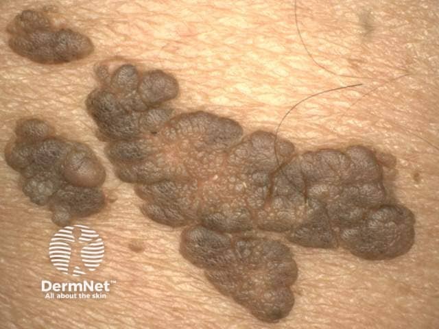
In the more common classical form of naevus lipomatosus superficialis, there are clusters of soft, yellow, skin-coloured lumpy lesions on the lower back, buttock and upper thigh, usually following a pattern of a single nerve distribution of that area (a zosteriform pattern). The nodules can be pedunculated or sessile with a smooth or wrinkled surface. They may be hairy or show comedo-like plugs.
In the solitary form, there is a single soft, yellow, skin-coloured subcutaneous swelling that can occur anywhere on the body.
The skin lesions are not associated with any symptoms.
What are the complications of naevus lipomatosus superficialis?
The main patient concern is cosmetic.
Naevus lipomatosus superficialis can rarely present with ulceration and necrosis.
How is naevus lipomatosus superficialis diagnosed?
Naevus lipomatosus superficialis is diagnosed by histopathological examination of a skin biopsy. Mature adipose cells are found in the dermis and are not connected to the subcutaneous tissue where fat cells usually reside.
What is the differential diagnosis of naevus lipomatosus superficialis?
The classical type of naevus lipomatosus superficialis may be misdiagnosed as:
The solitary type may be misdiagnosed as a:
- Fibroepithelial polyp (skin tag)
- Lipoma
- Accessory nipple
- Lymphatic malformation
- Haemangioma
- Neurofibroma
- Trichoepithelioma
- Cylindroma.
What is the treatment for naevus lipomatosus superficialis?
As naevus lipomatosus superficialis is a benign condition, it is best left untreated.
Surgical excision is the best option if treatment is needed for a cosmetic reason or if the lesion is ulcerated.
What is the outcome for naevus lipomatosus superficialis?
Excision of naevus lipomatosus superficialis is usually curative.
References
- Yap FB. Nevus lipomatosus superficialis. Singapore Med J. 2009; 50: e161–2. Available at: www.sma.org.sg/smj/5005/5005cr3.pdf (accessed 26 June 2019)
- Lima CDS, Issa MCA, Souza MB, Goes HFO, Santos TBPD, Vilar EAG. Nevus lipomatosus cutaneous superficialis. An Bras Dermatol 2017; 92: 711–13. DOI: 10.1590/abd1806-4841.20175217. PubMed Central
- Burns T, Breathnach S, Cox N, Griffiths C (eds). Rook's Textbook of Dermatology. 7th edn, Oxford: Blackwell Publishing, 2004.
On DermNet
- Naevus lipomatosus superficialis pathology
- Congenital naevi
- Skin lesions, tumours and cancers
- Hamartoma
Other websites
- Nevus Lipomatosus Superficialis — Dermatology Advisor