DermNet provides Google Translate, a free machine translation service. Note that this may not provide an exact translation in all languages

Translate

Nail matrix biopsy

Author: Dr Cliff Rosendahl, Associate Professor, School of Medicine, The University of Queensland, Australia, 2013. Reviewed by Prof Luc Thomas, Lyon 1 University, France. Updated by Dr Todd Gunson, Dermatologist, Auckland, New Zealand in July 2014.


toc-icon

What is the nail matrix?

The nail matrix consists of specialised cells that produce the nail plate. It is located at the end of the digit (finger or toe) under the the skin beyond the distal phalangeal joint. It can be seen protruding as a white half-moon shape at the base of some nails.  

What is nail matrix biopsy?

Nail matrix biopsy is a surgical procedure in which a tissue specimen is obtained from the nail matrix.

Why is nail matrix biopsy undertaken?

Nail matrix biopsy is undertaken to make or confirm a diagnosis of a disorder that is affecting the nail plate. The following list describes some conditions in which this procedure may be undertaken.

How is a nail biopsy done?

Nail matrix biopsy is usually undertaken under local anaesthetic.  Most often a digital block is performed, whereby the sensory nerves entering at the base of the affected finger or toe are numbed via an injection.

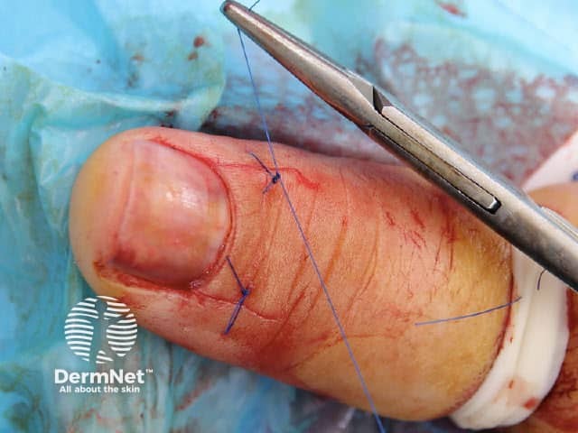
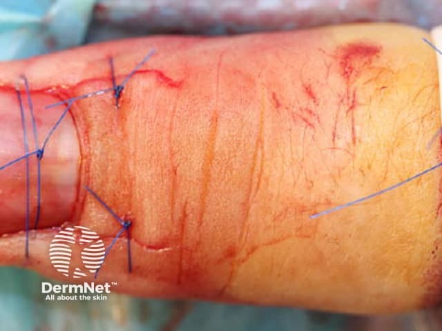
Various techniques are used to biopsy the nail matrix depending on the situation and the specialist's preference. The overlying nail plate may be removed for pathological examination, or replaced after the procedure. All techniques involve examining the matrix for the origin of the problem. It is important that adequate and appropriate sample is taken to make an accurate diagnosis, while minimising permanent damage/scarring to the matrix tissue.  

Techniques include:

  • Thin transverse incisional biopsy
  • Tangential (shave) biopsy
  • Punch biopsy

To minimise nail dystrophy, the biopsy should be performed on the distal matrix wherever possible, because this is responsible for producing the undersurface of the nail plate.   

Trap door technique

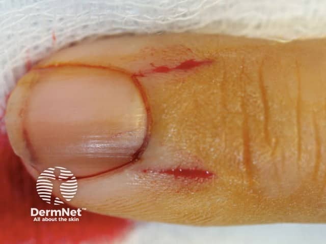
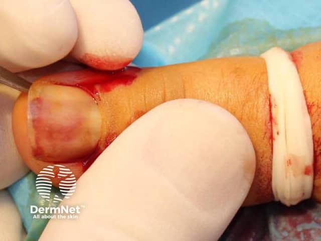
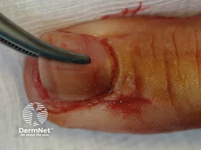
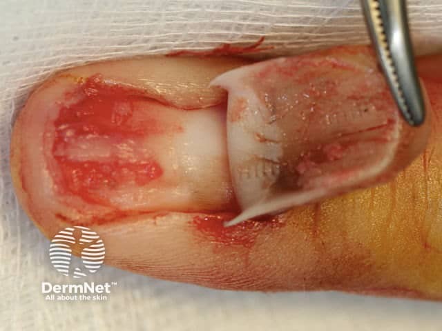
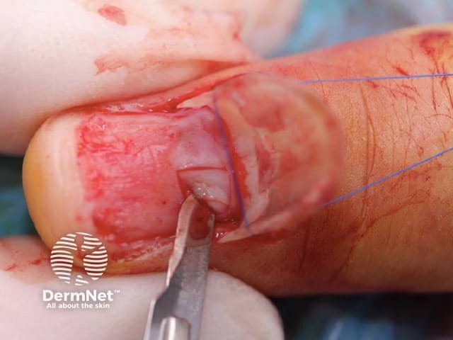
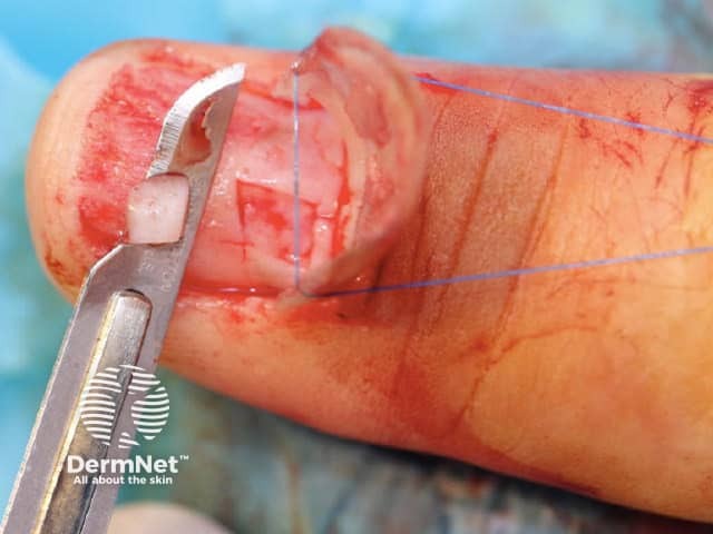
One technique is the “trap door” or “pop the bonnet” method used to biopsy of pigmented nail matrix lesions. This allows a direct view of the nail matrix permitting precise targeted biopsy of the lesion.

  • After local anaesthetic has been injected as a digital block using lignocaine without adrenaline, a tourniquet is applied to reduce bleeding. The time the tourniquet is applied is recorded. The proximal nail fold is completely cut through on each side approximately halfway back to the distal interphalangeal joint (figure 1).
  • The nail plate is separated from the nail bed with a Freer elevator, taking care to keep distal to the lunula (the half-moon) to avoid injuring the nail matrix (figure 2).
  • The nail plate is hinged up vertically like a car bonnet being opened (figure 3).
  • The nail bed has been injured by the procedure but the nail matrix is clearly and completely seen in pristine condition. Parallel lines of pigmentation can be seen in this case. The biopsy can be targeted to include a complete longitudinal sample of this portion of the nail matrix (figure 4).
  • The nail plate is retracted with a suture anchored on the tourniquet. The biopsy specimen is scored with a scalpel then shaved at least 1-mm thick. This is adequate for dermatopathological assessment if the lesion is thought to be thin. Extreme care is taken to avoid crushing the sample with forceps (figure 5).
  • The biopsy specimen is placed in formalin in a specimen container. The request form should carefully describe the lesion and the procedure. The specimen must be accurately labelled. It is recommended that the pathologist is directly contacted to ensure correct processing of the specimen (figure 6).
  • A suture is placed in each of the incisions (figure 7).
  • A suture is placed to hold the nail plate onto the nail bed (figure 8).
  • The tourniquet is removed and the tourniquet removal time should be recorded.
  • After removing the sutures a week later, the nail plate is vulnerable to “popping” and can be supported with tape.
Nail biopsy

What are the risks of nail biopsy?

Nail biopsy can lead to any of the usual complications from a surgical procedure, such as bleeding, infection, nerve damage, and scarring.

Scarring of the nail matrix is common and may result in permanent deformity of the nail plate.  Persistent paraesthesia (abnormal sensation) is one of the most common complications (7% in one study), likely due to damage to small nerves during the surgery. Overall, the complication rate following nail surgery performed by a specialist is low. 


References

  • Collins SC, Cordova K, Jellinek NJ. Alternatives to complete nail plate avulsion. J Am Acad Dermatol. 2008 Oct;59(4):619-26. doi: 10.1016/j.jaad.2008.05.039. Review. PubMed PMID: 18793936.

On DermNet

Books about skin diseases


Related information

Sign up to the newsletter