DermNet provides Google Translate, a free machine translation service. Note that this may not provide an exact translation in all languages

Translate

Melanonychia

Author: Ken Hiu-Kan Ip, Medical Student, University of Auckland, New Zealand; Chief Editor: Dr Amanda Oakley, Dermatologist, Hamilton, New Zealand; Copy Editor: Clare Morrison, June 2014. Updated by Dr Ebtisam Elghblawi and Dr Oakley in October 2017.


toc-icon
Table of contents
arrow-right-small

What is melanonychia?

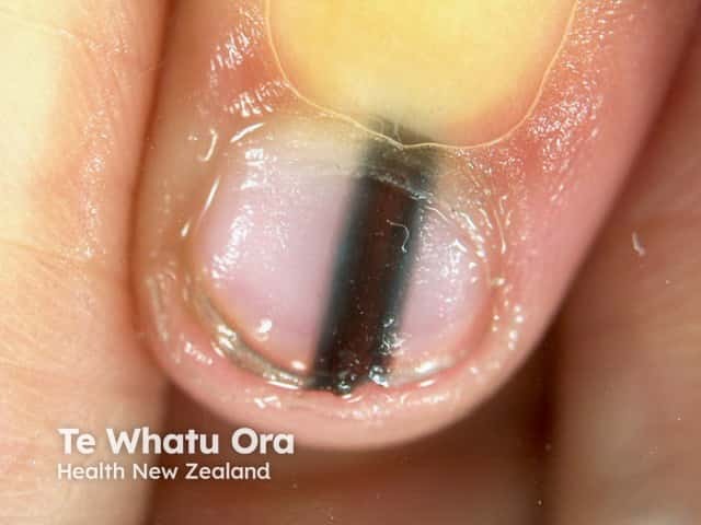
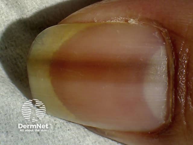
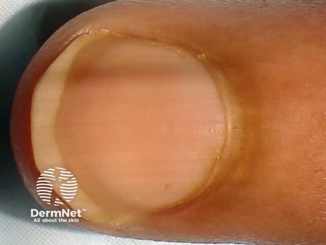
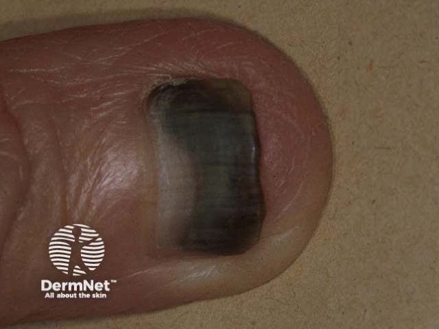
Melanonychia is brown or black discolouration of a nail. It may be diffuse or take the form of a longitudinal band.

Benign longitudinal melanonychia

See more images of melanonychia.

Who is at risk of melanonychia?

Melanonychia can present in individuals of all ages, including children, and affects both sexes equally. It is more prevalent in people with skin of colour, especially Fitzpatrick skin type V and VI.

  • Nearly all Afro-Caribbean people will develop black-brown pigmentation of the nails by the age of 50.
  • Melanonychia affects up to 20% of Japanese people.
  • White-skinned people are less commonly affected.

Melanonychia can also be associated with genetic disorders, injury, medications, nutritional deficiency, endocrine disease, connective tissue disease, inflammatory skin disease, a local tumour, or nail infection.

What causes melanonychia?

The nail plate is a hard, translucent structure made of keratin. It is not normally pigmented. Melanocytes typically lie dormant in the proximal nail matrix where the nail originates. Melanin is deposited into the growing nail when melanocytes are activated, resulting in a pigmented band — this is longitudinal melanonychia.

The deposition of melanin in the nail plate can result from 2 processes:

  • Melanocytic hyperplasia
  • Melanocytic activation.

Melanocytic hyperplasia

Melanocytic hyperplasia refers to an increased number of melanocytes within the nail matrix. This can represent a benign or malignant process.

Benign hyperplasia

Melanocytic naevi arise more commonly in children. Histologically there are nests of naevus cells.

Lentigines are seen more commonly in adults. Nests are absent.

Malignant hyperplasia

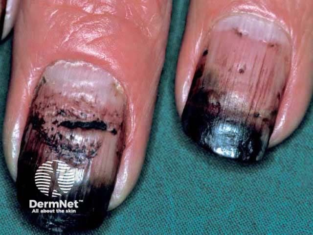
Melanoma of the nail unit most commonly affects the thumbs, index fingers and big toes.

Melanocytic activation

Melanocytic activation is an increase in the production and deposition of melanin into the nail cells (onychocytes), without an increase in the number of melanocytes. The causes of melanocytic activation are listed in the table below.

Melanonychia associated with melanocytic activation
Physiological (functional)

Racial variation

Pregnancy

Trauma

Nail biting, chewing, breaking and picking

Friction due to foot deformity or footwear

Inflammatory skin disease

Psoriasis

Acrodermatitis of Hallopeau

Lichen planus

Amyloidosis

Nonmelanocytic lesions

Intraepidermal carcinoma

Basal cell carcinoma

Viral wart

Nutritional deficiency Vitamin B12 or folate deficiency
Endocrine disorders

Addison disease

Cushing syndrome

Hyperthyroidism

Acromegaly

Another systemic disease

Haemosiderosis

Porphyria cutanea tarda

Human immunodeficiency virus infection (HIV)

Systemic lupus erythematosus

Systemic sclerosis

Syndromes

Laugier-Hunziker syndrome

Peutz-Jeghers syndrome

Touraine syndrome

Iatrogenic

Phototherapy

X-ray exposure; electron beam therapy

Medications

Chemotherapy agents (especially hydroxyurea, busulfan, bleomycin, adriamycin, doxorubicin, cyclophosphamide, 5-fluorouracil)

Anti-malarial therapy

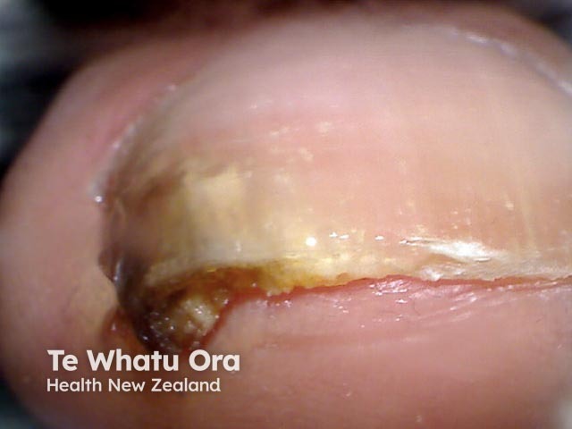
Pathogens can cause an irregular melanonychia as they stimulate inflammation activating melanocytes. For example:

External agents can stain the nails.

  • Hair dye
  • Henna
  • Enamel
  • Paint.

Discoloured nails

What are the possible complications of melanonychia?

Complications depend on the cause of melanonychia.

  • Subungual melanoma can result in metastasis and ultimately, the death of the patient. The prognosis for nail matrix melanoma is as a rule worse than melanoma in other sites.
  • Trauma, infection, and inflammatory disease can result in fissuring and splitting of the nails and unsightly or painful nail dystrophy.

What is the management of melanonychia?

Where melanonychia is attributed to a benign cause, no further treatment is necessary.

The management of melanoma of the nail unit requires complete excision of the tumour and may require amputation of the digit.

What is the outlook for patients with melanonychia?

Melanonychia tends to persist, except when it is related to medication — in which case it fades following its withdrawal.


References

  • Rook's Textbook of Dermatology. 4th edition. Eds: Rook A, Wilkinson DS, Ebling FJB, Champion RH, Burton JL. Blackwell Scientific Publications.
  • Mannava KA, et al. Longitudinal melanonychia: detection and management of nail melanoma. Hand Surgery 2013; 18: 133–9. PubMed
  • Jefferson J, Rich P. Melanonychia. Dermatol Res Pract 2012; 2012: 952186. PubMed
  • Haneke E, Baran R. Longitudinal melanonychia. Dermatol Surg 2001; 27: 580–4. PubMed
  • Benati E, Ribero S, Longo C, Piana S, Puig S, Carrera C, Cicero F, Kittler H, Deinlein T, Zalaudek I, Stolz W, Scope A, Pellacani G, Moscarella E, Piraccini BM, Starace M, Argenziano G. Clinical and dermoscopic clues to differentiate pigmented nail bands: an International Dermoscopy Society study. J Eur Acad Dermatol Venereol 2016. doi:10.1111/jdv.13991. Journal
  • Braun RP, Baran R, Le Gal FA, Dalle S, Ronger S, Pandolfi R, Gaide O, French LE, Laugier P, Saurat JH, Marghoob AA, Thomas L. Diagnosis and management of nail pigmentations. J Am Acad Dermatol 2007; 56: 835–47. PubMed

On DermNet

Books about skin diseases


Related information

Sign up to the newsletter