DermNet provides Google Translate, a free machine translation service. Note that this may not provide an exact translation in all languages

Translate

Dermoscopy

Dermoscopy of the nail

Created 2008.

Note: dermoscopic images in this course are nearly all at the same magnification; the full width of the image is equivalent to 12 millimetres on the patient.

Learning objectives

  • Recognise dermoscopic features of melanonychia

Introduction

Macroscopic views of nail pigmentation

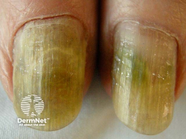
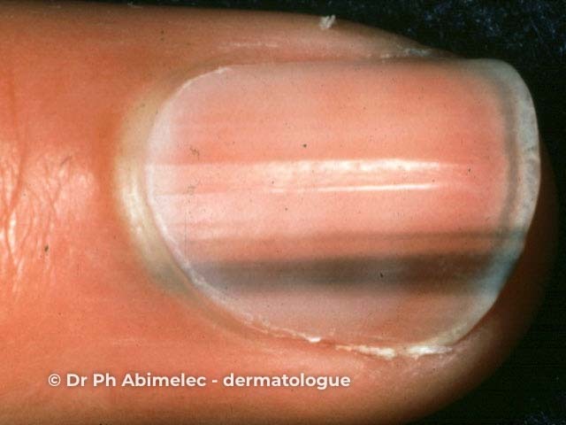
The nail plate is a semi-transparent keratinous structure and is not normally pigmented. It may appear discoloured because of pigment on top of the nail plate, within the nail plate or on top of the nail bed. Pigment may arise from the distal or proximal nail matrix.

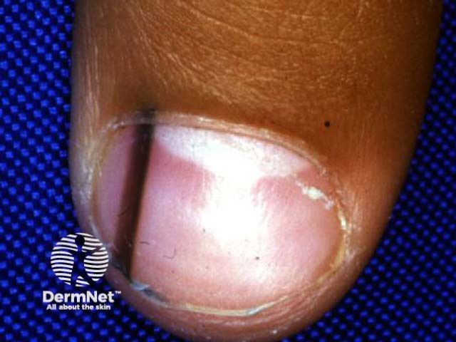
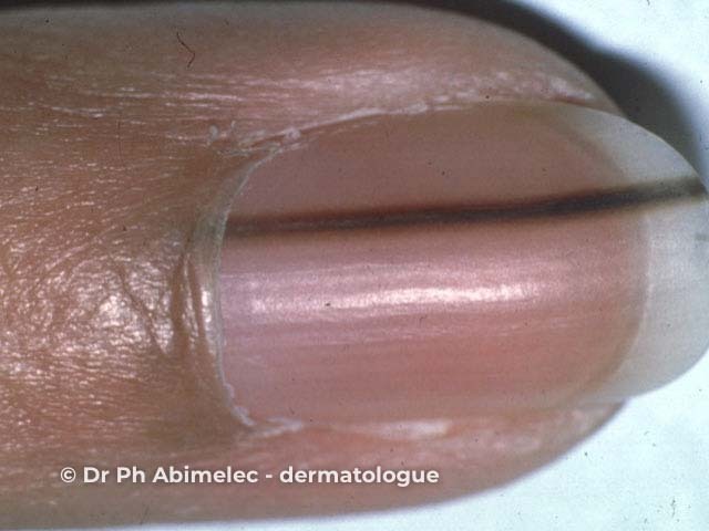
Longitudinal melanonychia is due to activation of melanocytes in the nail matrix. It is more common in darker skinned individuals and may affect one or several nails. The band may occasionally extend across the entire nail (total melanonychia). Transverse melanonychia is rare.

There are various causes of longitudinal melanonychia.

  • Exogenous pigment eg, silver nitrate, tobacco, henna
  • Ethnic pigmentation
  • Inflammatory skin disease (psoriasis, lichen planus)
  • Trauma (nail biting, friction from shoes, radiotherapy)
  • Infections (paronychia, onychomycosis especially when due to moulds; pigmentation is nonmelanocytic)
  • Drug reactions (hydroxyurea, antiretrovirals, antimalarials, metals)
  • Endocrine disease (Addison disease, Cushing syndrome)
  • Nonmelanocytic tumours (squamous cell carcinoma in situ, onychomatricoma, myxoid cyst, viral warts)
  • Melanocytic naevus of nail matrix
  • Lentigo / benign melanocytic hyperplasia
  • Malignant melanoma

Nails grow slowly, taking months to reach the distal edge, and longitudinal melanonychia reflects melanin deposition rather than the site of its production.

Melanonychia may be confused with discolouration due to blood spots. However, blood spots may also be seen in melanoma.

Nail plate dermoscopy

Macroscopic views of nail plate

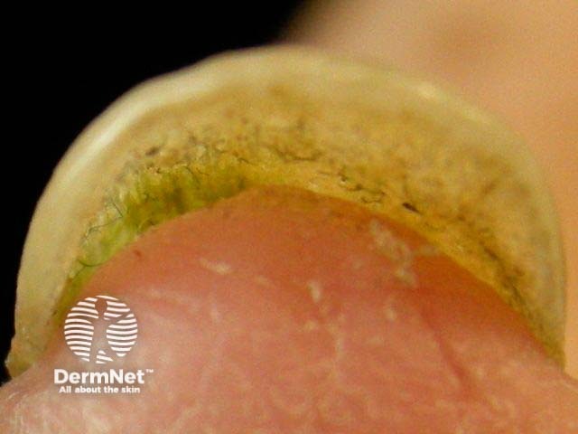
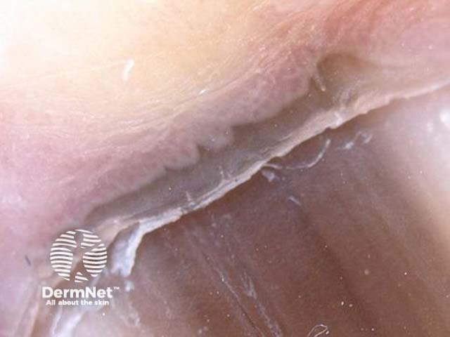
To improve the quality of the dermoscopic image, apply gel to the nail. Examine the nail plate from above as well as end-on (the free edge of the nail). Seen end-on:

  • Pigment in the top of the nail plate has its origin in the proximal matrix
  • Pigment at the bottom of the nail plate has its origin in the distal matrix or nail bed.

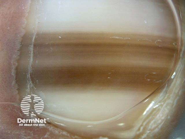
Dermoscopy is mainly used to assess pigmented streaks or bands, which usually extend from the proximal nail fold adjacent to the cuticle to the distal edge. Several structures have been described.

  • Blood spots (subungual haemorrhage)
  • Light to dark brown background colour or bands
  • Regular thin lines (melanocytic hyperplasia)
  • Irregular lines (melanoma)
  • Grey background and thin grey lines (epithelial melanin)
  • Micro-Hutchinson sign (cuticular pigmentation not easily seen by naked eye examination)
  • Microscopic grooves
  • Granular inclusions (tiny grey or brown dots indicating melanocytic origin)

Benign melanonychia

Melanocytic naevus

Melanonychia due to melanocytic naevus

Melanocytic naevus of the nail apparatus is characterised by:

  • Regular parallel lines
  • Brown background
  • Granular inclusions

Dark pigmented bands may result in the pseudo-Hutchinson sign, in which the pigmentation is visible through the transparent nail fold.

Epithelial melanin

Melanonychia due to epithelial melanin

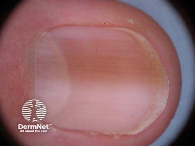
Epithelial melanin results in ethnic-type pigmentation, lentigo, and drug-induced pigmentation. The resulting melanonychia is characterised by:

  • Homogeneous longitudinal thin grey lines
  • Light brown to dark grey background colour.

Benign melanonychia due to epithelial melanin may affect multiple nails, particularly in individuals with skin phototype 5 or 6. They are more often observed on fingernails than toenails. They may be seen as part of Laugier-Hunziker syndrome or Peutz-Jeghers syndrome.

Congenital melanocytic naevi

Congenital melanocytic naevi of the nail matrix are uncommon but may appear alarming. Features may include:

  • Involvement of nail fold (Hutchinson sign) and hyponychium, often with parallel furrow pattern or a variant
  • Variation in the width of the longitudinal pigmented band, often triangular
  • Black dots
  • Thinning and fissuring of the nail plate
  • Fading and disappearance of pigmentation

Malignant melanoma

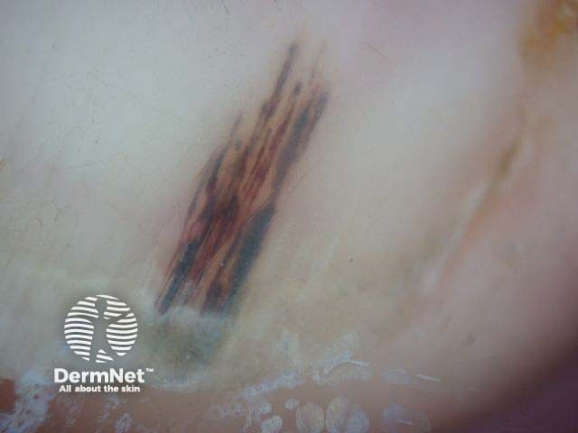
Melanoma should be considered if pigmentation affects a single nail, especially if it is of recent origin in an adult. Thumb nail and great toenail are most often affected. Melanoma may affect the nail bed (subungual) or matrix (pigment within nail plate).

Nail matrix melanoma

Dermoscopic features of nail matrix melanoma include:

  • Longitudinal brown to black parallel lines with irregular colouration, spacing, or thickness
  • Disruption of parallelism, for example, pigmentation increases in width proximally
  • Brown background
  • Hutchinson sign: pigmentation of nail fold
  • Nail plate fissuring or destruction

These features are not entirely reliable and should not be used as a substitute for biopsy if there is any doubt about the origin of longitudinal melanonychia.

Subungual melanoma

Nail bed melanoma

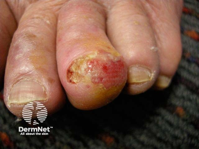
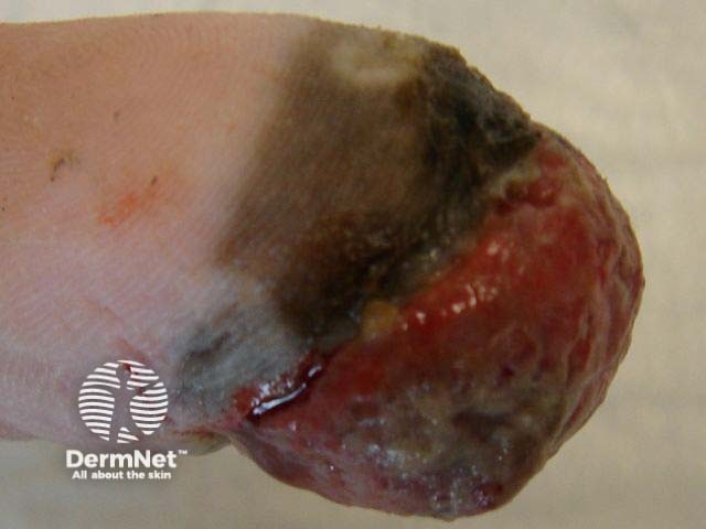
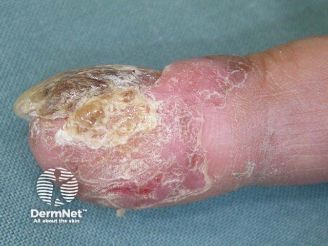
Nail bed melanoma or subungual melanoma results in a red, brown or black nodule under the nail plate, which ulcerates and bleeds. It may resemble pyogenic granuloma.

Subungual haemorrhage

Subungual haemorrhage

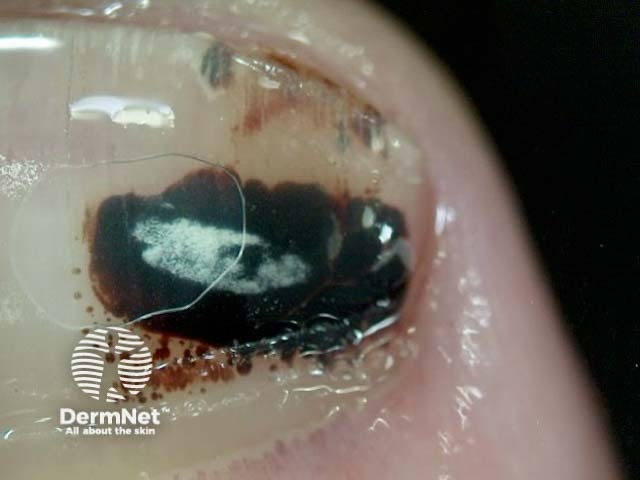
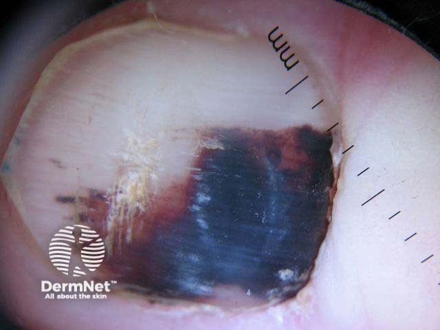
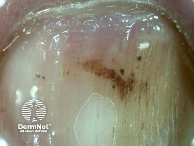
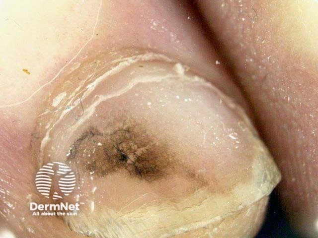
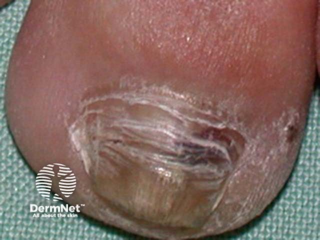
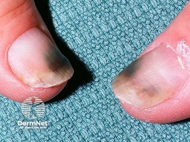
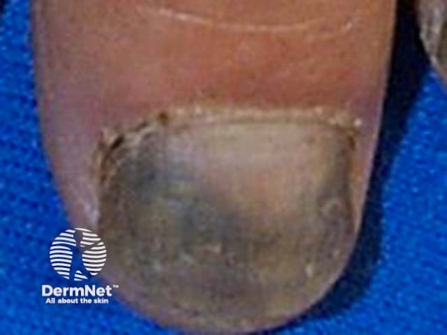
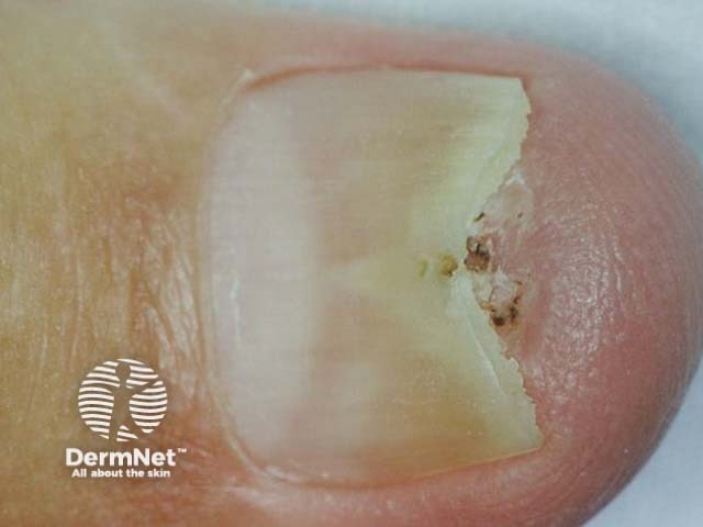
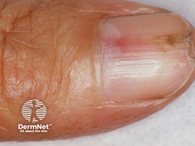
Subungual haemorrhage is the most common cause for pigmentation of the nail; patients often deny trauma and may be unable to give a clear history. The presence of red to bluish-black blood spots on dermoscopy is helpful.

Blood spots are well-circumscribed dots, globules or blotches; they may be red, purple, blue, brown or black. The proximal edge tends to be rounded and well circumscribed, whereas the distal edge is more likely to show parallel linear structures.

Blood spots sometimes grow out more slowly than the nail because they are under the nail plate rather than incorporated within it. Treat recurrent haemorrhage with suspicion; a tumour may be responsible.

Nonpigmented lesions

Macroscopic views of nonpigmented lesions

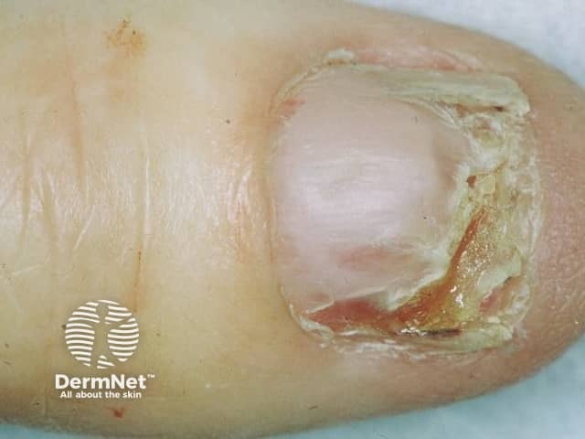
Tumours of the nail unit may be nonpigmented, when diagnosis is frequently delayed. They include benign and malignant lesions.

  • Haemangioma
  • Onychomatricoma
  • Glomus tumour
  • Invasive or in-situ squamous cell carcinoma
  • Amelanotic melanoma

Dermoscopic features to be evaluated include:

  • Remnants of pigmentation in melanoma
  • Atypical blood vessels in melanoma
  • Microhaemorrhage in melanoma, squamous cell carcinoma, onychomatricoma
  • Longitudinal erythronychia in haemangioma, onychomatricoma, glomus tumour
  • Yellow or white nail plate, subungual hyperkeratosis in squamous cell carcinoma, onychomatricoma, exostosis
  • Triangular onycholysis in squamous cell carcinoma,, glomus tumour
  • Blue spots in blue naevus, glomus tumour, melanoma

Activity

What are the indications for nail biopsy? [See Braun et al's article below.]


Related information

References

  • S. Ronger, S. Touzet and C. Ligeron et al., Dermatoscopic examination of nail pigmentation, Arch Dermatol 2002: 138; 1327–1333
  • Tosti A. Dermoscopy Allows Better Management of Nail Pigmentation. Arch Dermatol. 2002;138:1369-1370.
  • Tosti A, Piraccini BM, de Farias DC. Dealing with Melanonychia. Seminars in Cutaneous Medicine and Surgery 2009; 28:49-54
  • Braun RP, Baran R, Le Gal FA, Dalle F, Ronger S, Pandolfi S, Gaide O, French LE, Laugier P, Saurat JH, Marghoob AA, Thomas L. Diagnosis and management of nail pigmentations. Journal of the American Academy of Dermatology 2007; 56:835-847.

On DermNet NZ


Books about skin diseases

Text: Miiskin

Sign up to the newsletter