DermNet provides Google Translate, a free machine translation service. Note that this may not provide an exact translation in all languages

Translate

Dermoscopy

Dermatoscopic-histologic correlation

Created 2008.

Learning objectives

  • Describe histopathological correlates of dermatoscopic features of pigmented and non-pigmented skin lesions

Introduction

This section on dermatoscopic-histologic correlation is not intended to be a comprehensive overview of dermatoscopic diagnosis, which is covered elsewhere on this site. The terminology used is, however, similar and is based on the monograph by Kittler H et al.[1], on modified pattern analysis. Unless otherwise indicated, the diagrams in this section are reproduced from that monograph with the kind permission of the authors.

There is a close correlation between the patterns and features observed dermatoscopically and the histological features of biopsied or excised skin lesions, particularly in the case of pigmented lesions. With experience it is possible to predict the features that will be observed histopathologically based on the dermatoscopic examination. This correlation is important for both clinician and pathologist to understand as the two diagnostic modalities provide complementary information when used in the diagnosis of pigmented skin lesions [2]. This reflects the different ways in which they view the same lesion. Dermatoscopy provides an overview of the lesion viewed parallel to the skin surface, akin to an aerial photograph of a landscape, whereas histopathology provides a detailed assessment of a small part of the lesion that is viewed perpendicular to the skin surface, akin to a photograph from ground level of a hillside within the overall landscape. Correlation is essential to ensure that the pathologist has viewed the parts of a lesion in which specific dermatoscopic features of interest or concern have been noted. Using the analogy above one may not appreciate from a ground level photograph what is beyond the hill you are photographing whereas this may be obvious in an aerial photograph.

It is essential, therefore, that any pathology report issued on a skin lesion on which dermatoscopy has been performed should detail the histologic correlate of the dermatoscopic features noted. It is equally important that a dermatoscopist should be satisfied that the pathologist has provided an adequate explanation for the features they observed. If there is any doubt a phone call to clarify the situation and perhaps examination of deeper levels by the pathologist is indicated.

Dermatoscopic–histologic correlation of pigmented skin lesions

The dermatoscopic assessment of pigmented lesions using pattern analysis involves assessment of colour and pattern and the identification of clues to a specific diagnosis. Colour and pattern are used to assess the symmetry of a lesion. Each of these components of the assessment correlates with the histopathology.

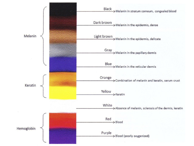
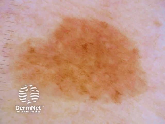
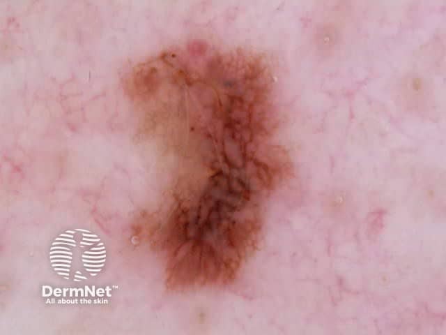
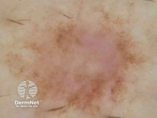
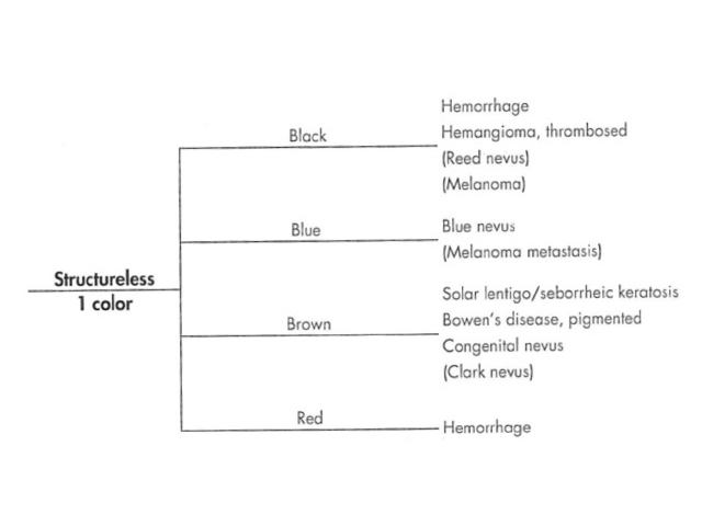
Colours: The colours observed dermatoscopically correlate with the nature of the pigment / substance and, in the case of melanin, the depth of the pigment observed histopathologically (Figure 1).

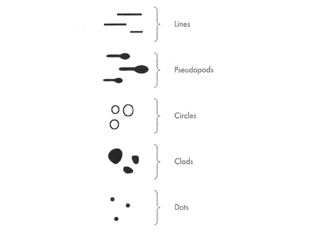
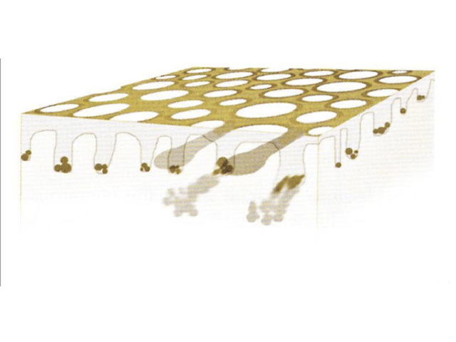
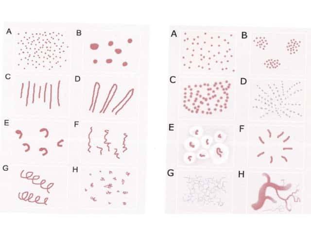
Elements: Dermatoscopic patterns are formed by the repetition of, or absence of a repetitive pattern of, one of five basic elements. These are lines, pseudopods, circles, clods and dots (Figure 2). The absence of a dominant repetitive pattern of one of these elements typifies the structureless pattern. You can click on the images for a larger view.

Colours and elements in dermatoscopy

  • Figure 1. Diagram. Colours in dermatoscopy. From Kittler H et al [1].
  • Figure 2. Diagram. Five basic elements. Multiple repetitions of each can constitute a pattern. From Kittler et al [1].

Patterns of lines

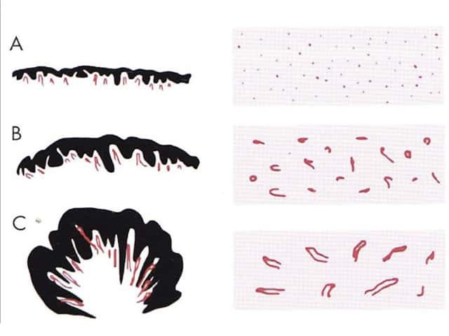
There are five dermatoscopic patterns of lines. These are reticular, branched, parallel, radial and curved (Figure 3).

Patterns of lines on dermatoscopy

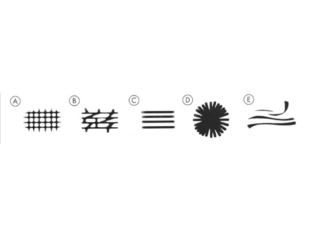
Figure 3. Diagram. Five possible patterns of lines. From Kittler et al [1].

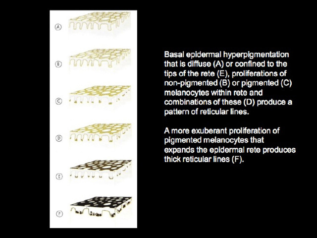
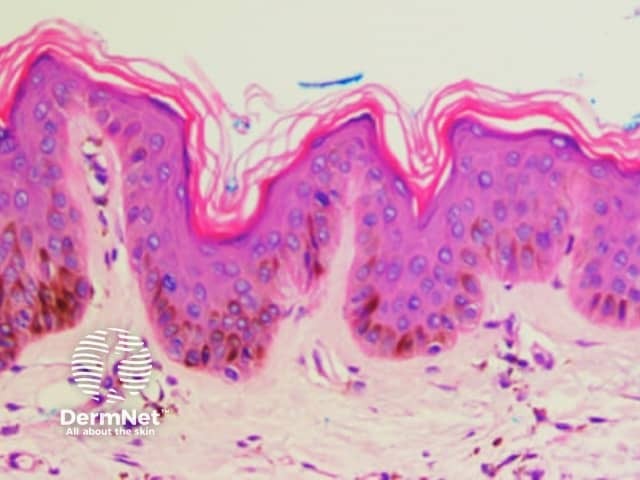
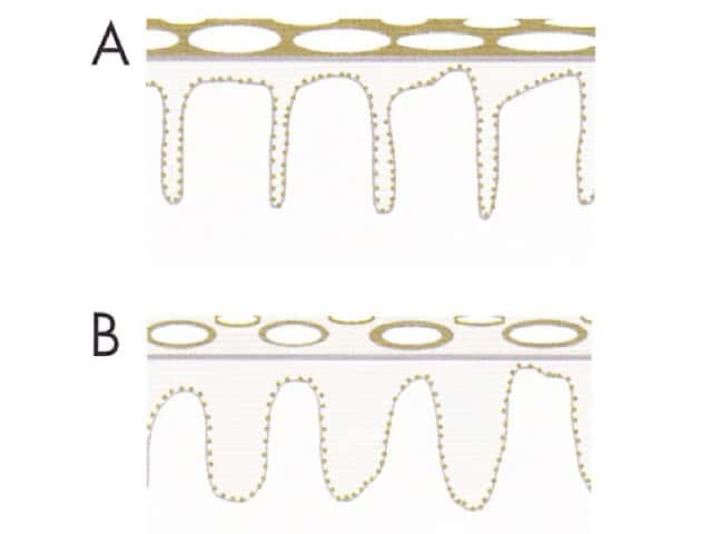
Apart from radial lines, dermatoscopic patterns of lines reflect either the normal interdigitation of epidermal rete ridges (downgrowths) and dermal papillae (upward indentations, Figure 4a ); exaggerations, caused by acanthotic keratinocytic proliferations (such as seborrhoeic keratosis); or distortions of that pattern (such as broadened network due to exuberant melanocytic proliferation expanding epidermal rete ridges). Site specific variations in the anatomy of the dermoepidermal junction account for certain patterns, such as parallel lines on acral sites, or variations in the patterns seen in similar lesions in different anatomic sites.

Pathology of normal skin.

  • Figure 4a. Histopathology of skin. Normal interdigitation of epidermal rete (arrows) and dermal papillae (stars).

Radial lines
Radial lines are the dermatoscopic manifestation of peripheral extensions of pigmented lesional cells such as pigmented basal cell carcinoma or elongate nests of pigmented melanocytes in melanocytic proliferations. As radial lines are viewed parallel to the epidermal surface they are not evident in basal cell carcinoma sectioned perpendicular to the epidermal surface for histolological examination, precluding specific correlation. In melanocytic lesions they comprise elongate nests of melanocytes similar to pseudopods on histology. As such, the histologic correlation of radial lines will not be discussed further.

Reticular and branched lines
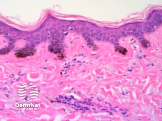
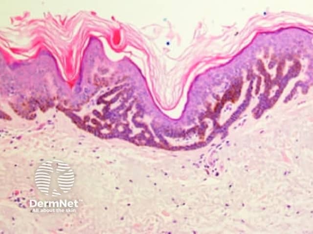
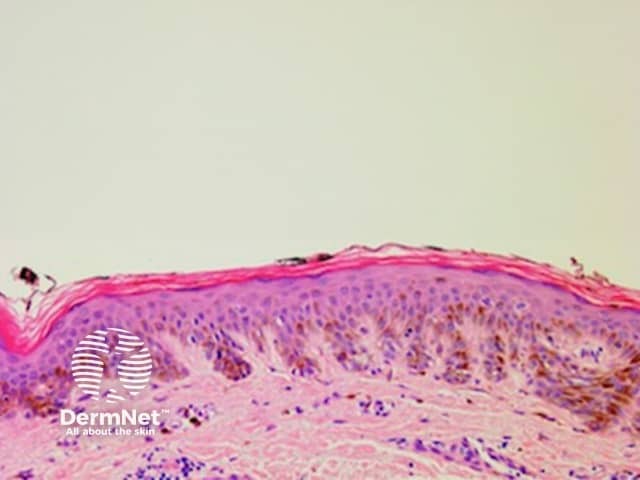
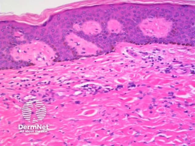
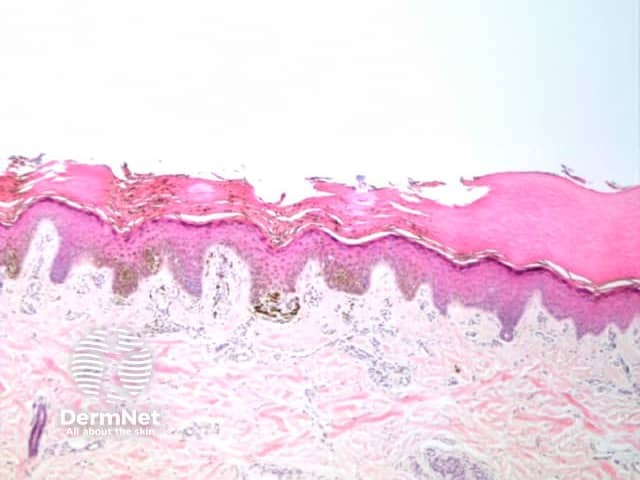
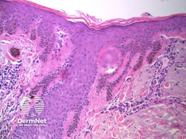
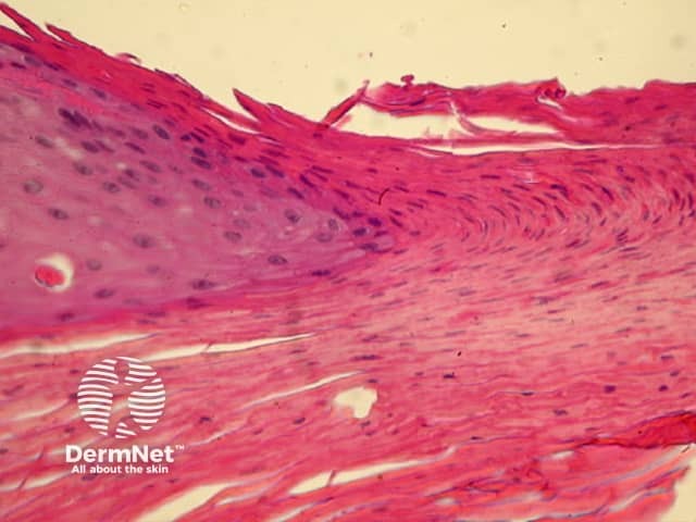
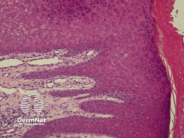
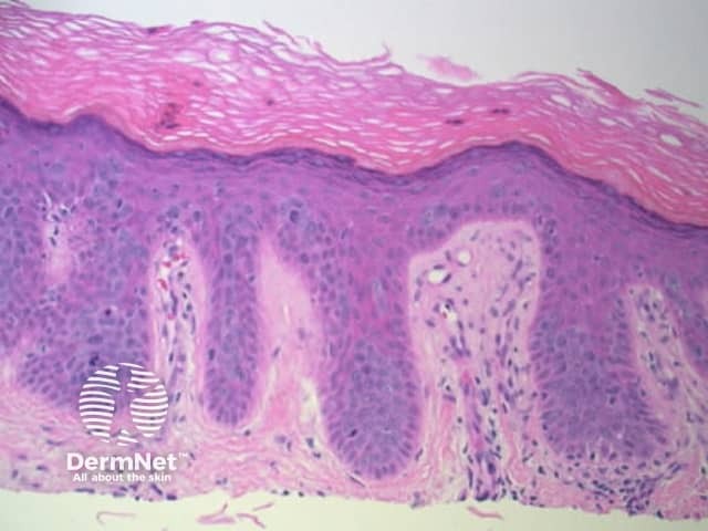
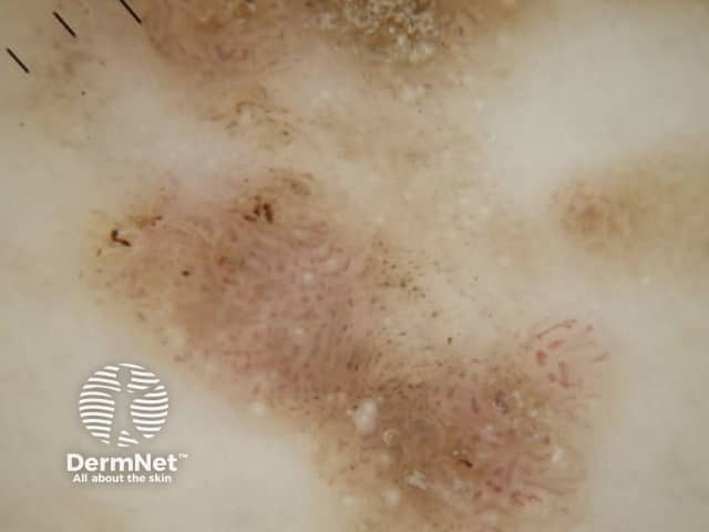
Reticular lines are straight and intersect at regular intervals at right angles. Branched lines are also straight but intersect at irregular intervals and not at right angles. The distinction is not always clear cut and rarely of great importance. These dermatoscopic patterns are seen in lesions in which there is basal epidermal hyperpigmentation and / or a proliferation of pigmented melanocytes preferentially involving the epidermal rete ridges with preservation of the epidermal architecture (Figure 5).

Reticular / branched lines

  • Figure 5. Diagram. The histologic basis for reticular / branched lines. From Kittler et al [1].

Curved lines
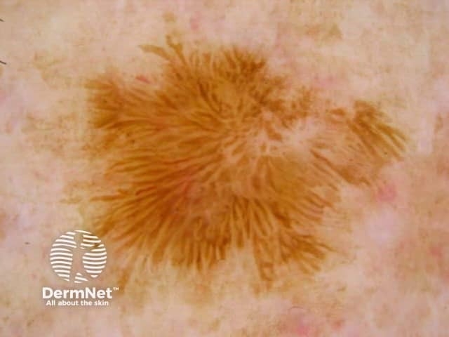
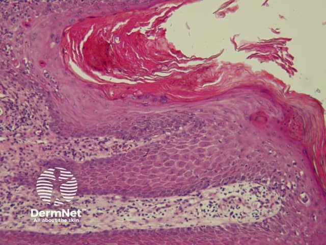
Curved lines are seen in dermatoscopy of pigmented keratinocytic proliferations that are either acanthotic, such as seborrhoeic keratosis, or in specific locations, such as close to mucocutaneous junctions, as with some labial and genital melanotic macules.

Lesions characterized by reticular / branched / curved lines

  1. Melanotic macules

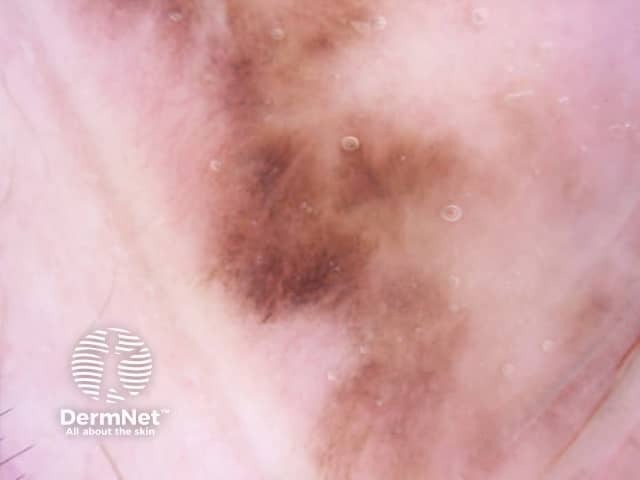
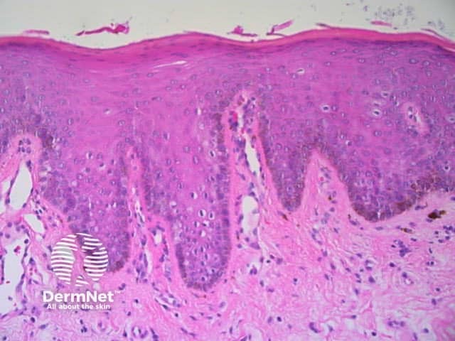
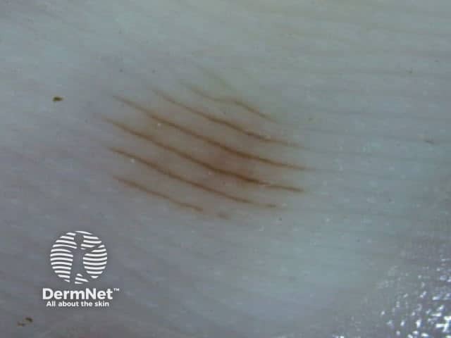
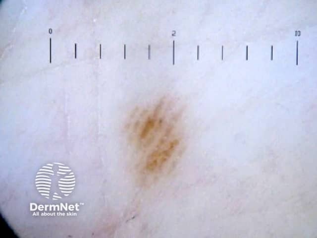
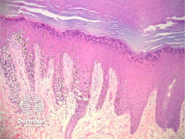
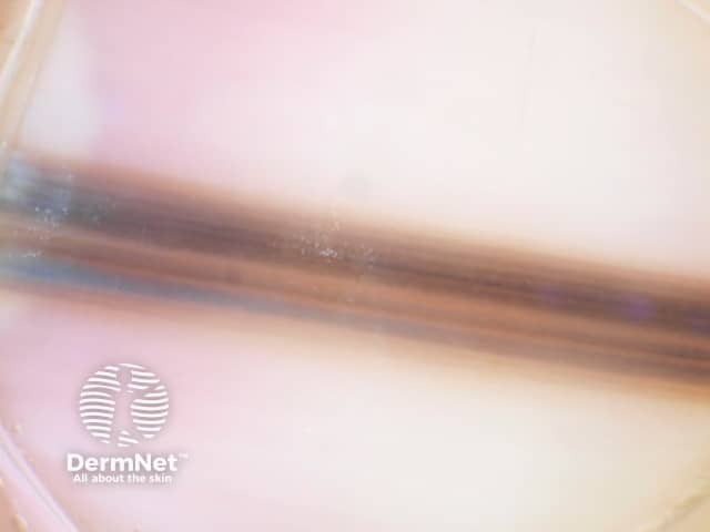
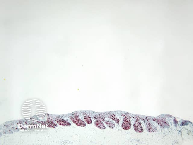
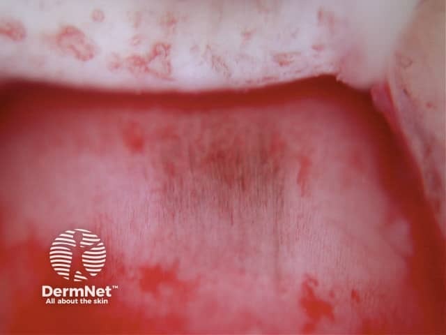
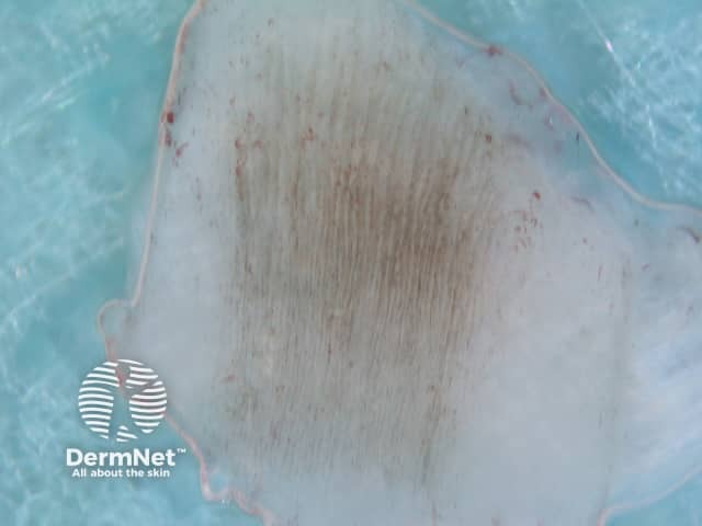
The dermatoscopic features of melanotic macules vary with site reflecting the differences in the anatomy of the dermoepidermal junction in normal skin (ink spot lentigo, Figures 6a, 6b) and mucocutaneous transition zones (Figures 7a and 7b). The histology is, however, reasonably similar irrespective of site (Figure 8).

Dermatoscopic images of ink spot lentigo

  • Figure 6a. Dermatoscopy of ink spot lentigo. Reticular dark brown lines that end abruptly at the margin of the lesion. Courtesy Dr Alan Cameron.
  • Figure 6b. Dermatoscopy of ink spot lentigo. Branched dark brown lines that end abruptly at the margin of the lesion. Courtesy Dr Cliff Rosendahl.

Dermatoscopic images of mucosal melanotic macules

  • Figure 7a. Dermatoscopic images of labial (7a) melanotic macule. These lesions can show 3 different patterns on dermatoscopy: curved parallel lines, circles or structureless. The first two likely represent the unique configuration of the dermoepidermal junction in mucocutaneous transition zones, whereas the latter reflects the presence of flattened rete in some lesions. Courtesy Dr Cliff Rosendahl.
  • Figure 7b. Dermatoscopic images of genital melanotic macule. Appearance of genital melanotic macules is similar to labial melanotic macules. Courtesy Dr Cliff Rosendahl.

Histology of melanotic macule

  • Figure 8. Histology of melanotic macule. This is reasonably similar in skin and mucosa, and shows epidermal acanthosis with pigmentation of the basal layer at the tips of the rete ridges and often underlying superficial dermal melanophages. This corresponds to the situation in Figure 5(E).
  1. Solar lentigo

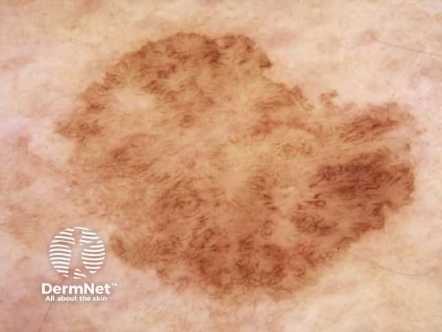
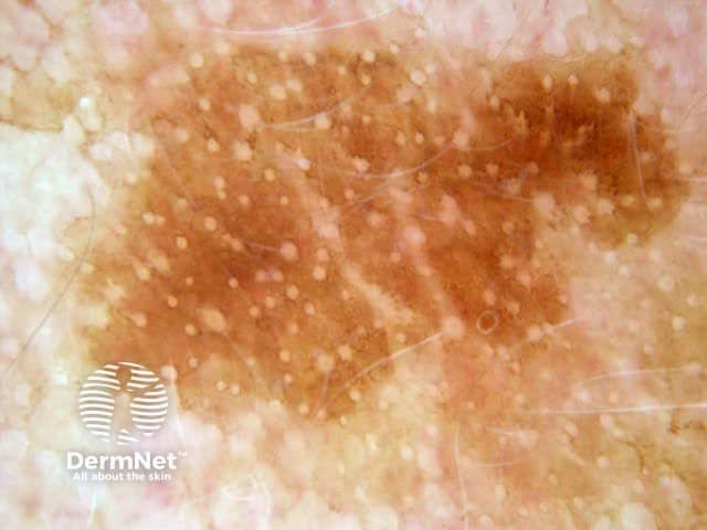
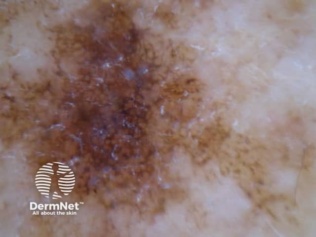
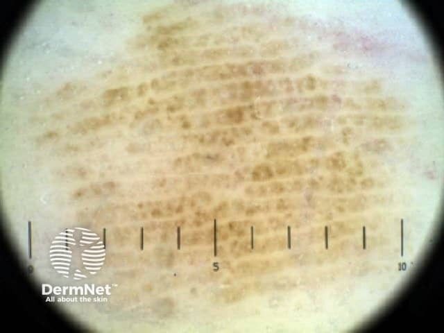
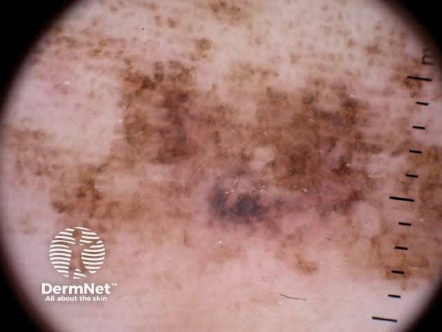
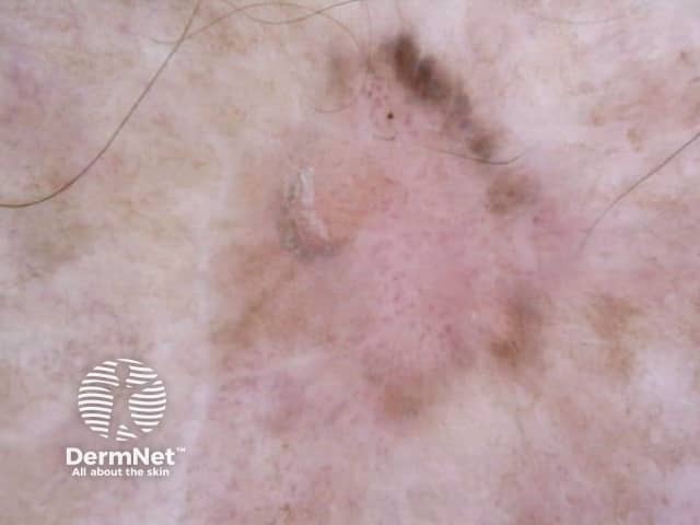
The dermatoscopic features observed in solar lentigo depend on the site of the lesion (Figures 9, 10 and 11).

Dermatoscopy of solar lentigines

  • Figure 9. Dermatoscopy of solar lentigo from trunk. These lesions typically show a pattern of reticular or curved lines, sometimes with superimposed circles. Courtesy Dr Alan Cameron
  • Figure 10. Dermatoscopy of solar lentigo from the face. These typically show a structureless pattern with hypopigmented follicular openings, as in this case, or regularly spaced circles. Lines are usually inconspicious due to the flattened dermoepidermal junction in sun damaged facial skin. Courtesy Dr Alan Cameron.
  • Figure 11. Dermatoscopy of solar lentigo from forearm. In this site a structureless dermatoscopic pattern and/or dots is typical. Courtesy Dr Alan Cameron.

Histology of solar lentigo

  • Figure 12. Histopathology of prototypic solar lentigo. Bulbous pigmented epidermal rete ridges are present. These are manifest as a pattern of lines dermatoscopically. Lines are not often apparent in facial lesions, due to flattening of the dermoepidermal junction, and in forearm lesions where the downgrowths are typically less even and the dermoepidermal junction may be more effaced due to chronic sun damage.
  1. Seborrhoeic keratosis

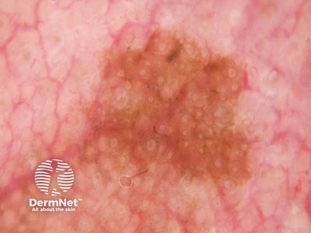
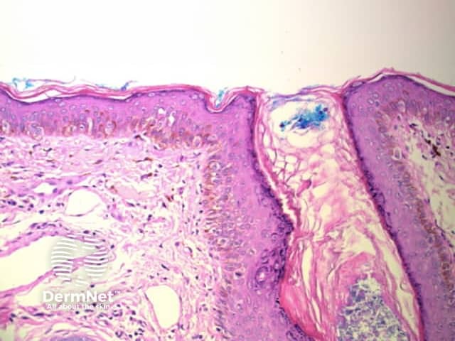
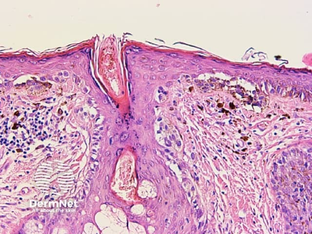
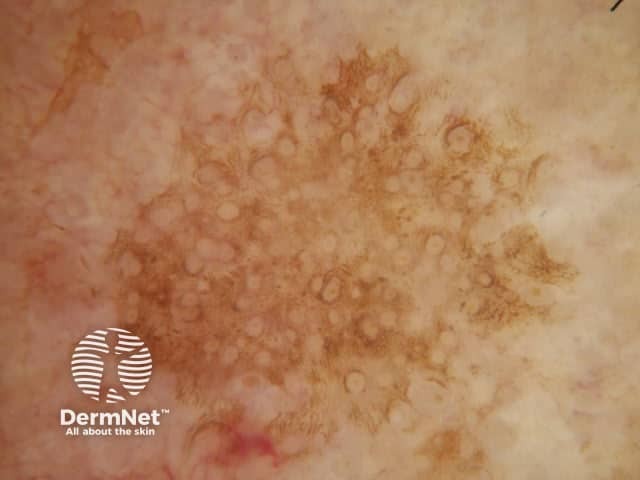
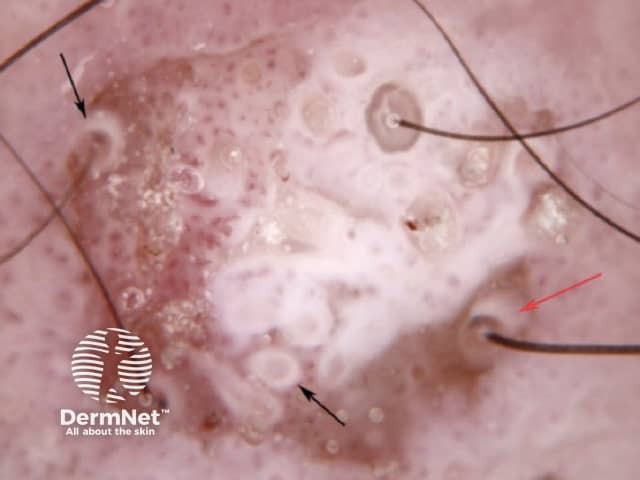
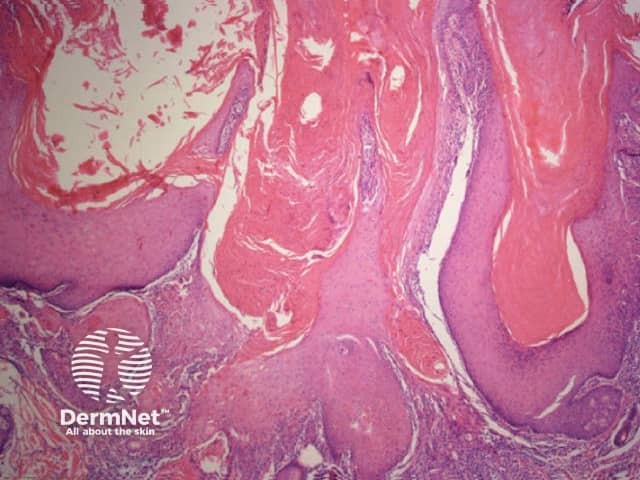
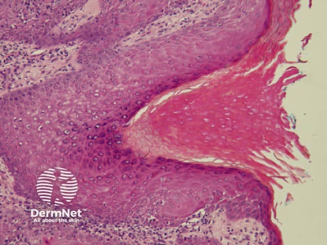
Seborrhoeic keratoses are amongst the most variable lesions on dermatoscopy, which is reflected in their protean histological appearances (Figure 15). Aside from the pseudopod pattern, any pattern or colour can be found. Flat seborrhoeic keratoses appear similar to solar lentigo on dermatoscopy. With early epidermal acanthosis thin curved lines (Figure 13) and circles become manifest, whilst with advanced acanthosis thick curved lines (Figure 14) and clods typically predominate. White clods are due to keratin under the stratum corneum. Orthokeratotic loosely laminated surface keratin appears yellow. Crypts fill with discoloured keratin and may appear various colours on dermatoscopy including brown and orange (Figure 15).

Dermatoscopy of seborrhoeic keratoses

  • Figure 13. Dermatoscopy of seborrhoeic keratosis. Pattern of thin curved lines. Courtesy Dr Cliff Rosendahl.
  • Figure 14. Dermatoscopy of seborrhoeic keratosis. Pattern of thick curved lines. Courtesy Dr Alan Cameron.
  • Figure 15. Dermatoscopy of seborrhoeic keratosis. Pattern of orange and white clods. Courtesy Dr Alan Cameron.

Histology of seborrhoeic keratoses

  • Figure 15a. Histopathology of flat seborrhoeic keratosis. This may closely resemble solar lentigo on dermatoscopy.
  • Figure 15b. Histopathology of flat seborrhoeic keratosis. Thin curved lines seen on dermatoscopy correspond to early acanthosis.
  • Figure 15c. Histopathology of thicker seborrhoeic keratosis. Thick curved lines seen on dermatoscopy are due to advanced epidermal acanthosis. Horn pseudocyst is the white clod seen on dermatoscopy.
  1. Dysplastic (Clark) naevus

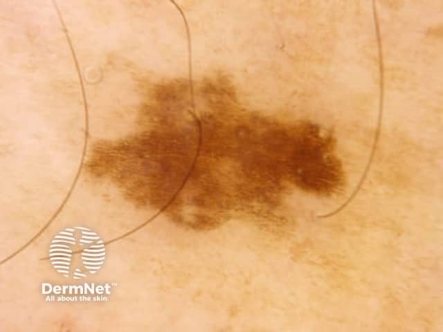
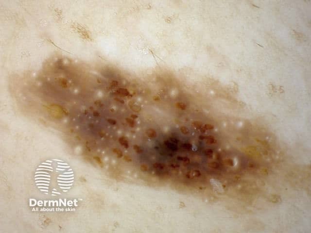
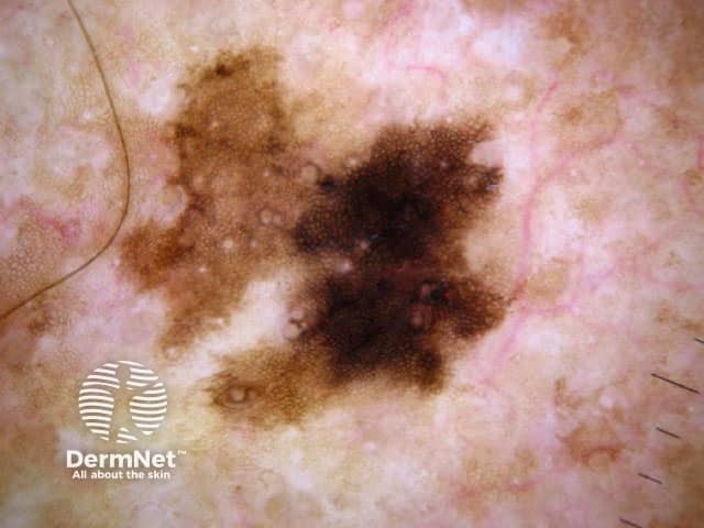
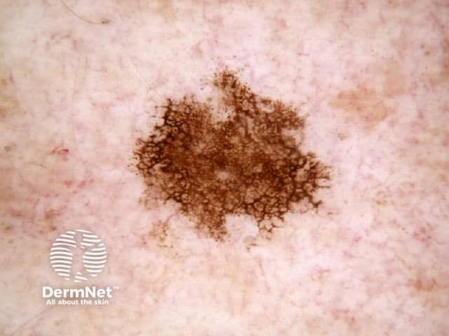
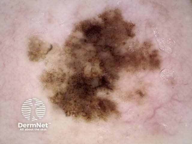
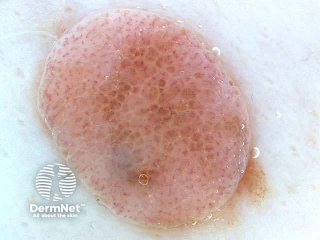
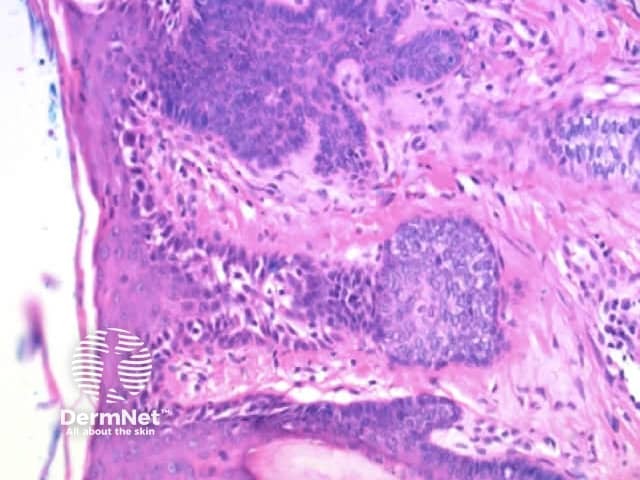
Both lentiginous and dysplastic naevi (Clark naevi) typically show a pattern of reticular lines on dermatoscopy, or less often circles (see later), that may be combined with dots, clods or structureless areas, usually in a symmetrical arrangement. Rarely only dots or clods or structureless areas are present.

Dysplastic (Clark) naevus

  • Figure 16. Dermatoscopy of dysplastic (Clark) naevus. Courtesy Dr Peter Bourne
  • Figure 17. Histopathology of dysplastic (Clark) naevus. Elongated pigmented rete ridges in association with a lentiginous and nested melanocytic proliferation showing some variability in melanocyte size. The delicate elongate rete are a consistent finding that manifest as reticular lines on dermatoscopy.

Thick lines

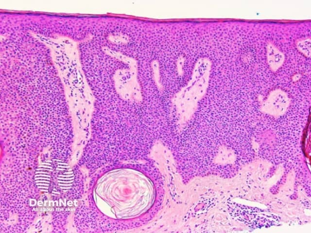
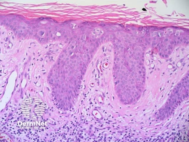
Thick lines are defined in dermatoscopy as those in which the lines are as thick, or thicker, than the spaces between them. Thick reticular or branched lines can be seen when an exuberant proliferation of pigmented melanocytes expands the epidermal rete ridges (Figure 18a,b). Thick curved lines are seen when a pigmented keratinocytic lesion produces acanthosis with thick downgrowths of pigmented epithelium as in some seborrhoeic keratoses (see previous). Thick parallel lines are discussed seperately later.

Histology of melanoma that has thick lines on dermatoscopy

  • Figure 18a. Dermoscopy of in situ melanoma. Nests of atypical melanocytes bridging the rete ridges results in thick lines.
  • Figure 18b. Dermoscopy of invasive melanoma with thick lines.
  • Figure 18c. Histology of invasive melanoma (not that in 18a). More exuberant melanocytic proliferation can expand epidermal rete and produce thick lines. Expanded rete are seen in the in-situ component.

Reticular / branched lines combined with other patterns

Reticular and branched lines are commonly seen in association with other dermatoscopic patterns, either symmetrically or asymmetrically. The combinations with dots and clods reflects the presence of those things that cause those patterns (eg large nests of pigmented melanocytes giving clods) in conjunction with elongate pigmented rete.

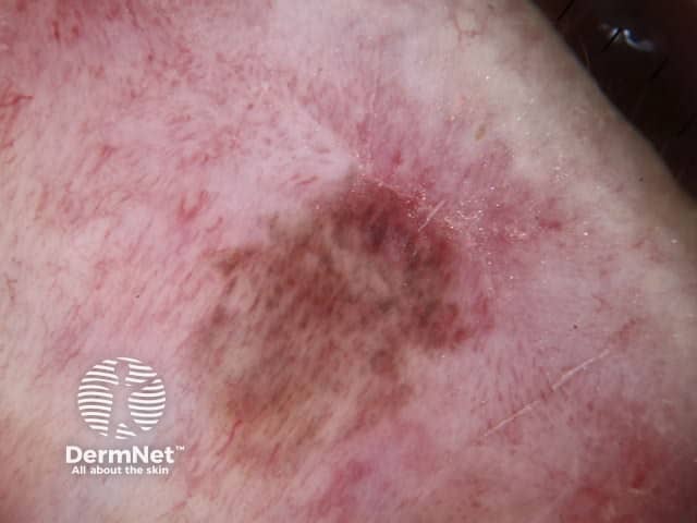
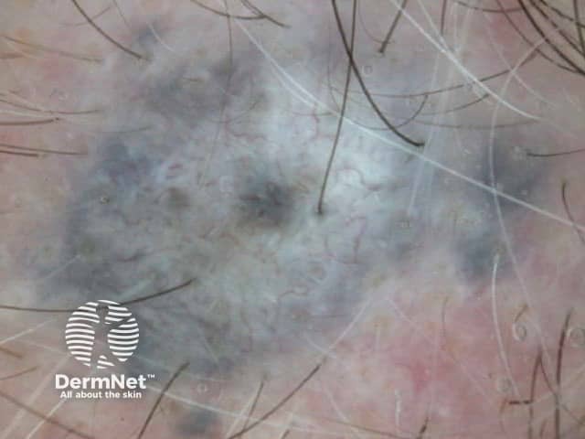
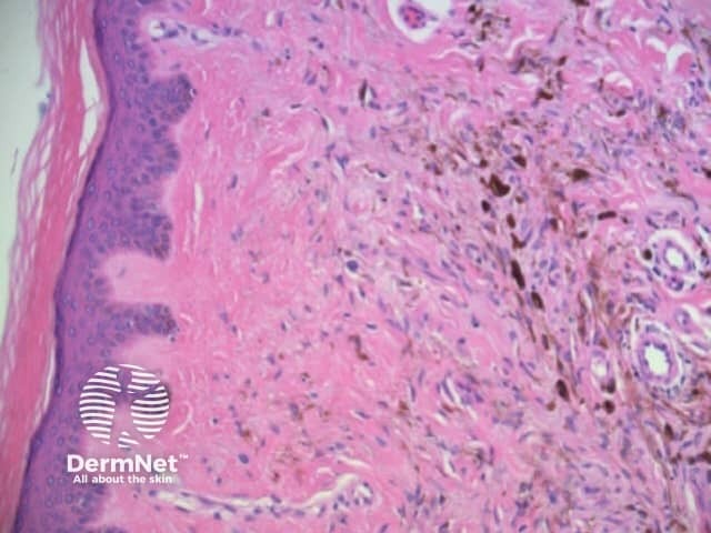
The combination of dermatoscopic structureless and reticular patterns may be due to marked pigmentation of lesional cells obscuring the pattern of lines and / or loss of the epidermal rete in part of the lesion. The former can be seen centrally in some dysplastic naevi, centrally or eccentrically in combined (true and blue) naevi and eccentrically in some melanomas. The latter may be seen with epidermal remodelling in melanoma (Figure 19a,b,c), past regression of melanocytic proliferations or due to an underlying expansile dermal lesion (Figures 20-22). The colour of the structureless zone depends on the level of the pigment within the skin: it may be white, if due to dermal fibrosis; skin coloured, if associated with epidermal remodelling in association with a proliferation of non-pigmented melanocytes; and black, brown, grey or blue, if due to heavy melanin pigmentation.

Melanoma with structureless zones on dermatoscopy

  • Figure 19a. Dermatoscopy of melanoma reported to be in situ reveals lines and pigmented and blue-grey structureless zones.
  • Figure 19b. Dermatoscopy reveals irregularly distributed lines, and pigmented and whitish structureless zones. Histology of this melanoma in situ showed marked fibrosis, melanin in melanophages, and regression.
  • Figure 19. Histology of lentiginous level 1 melanoma (melanoma in situ). The effacement of epidermal rete on the right side of this image in association with a proliferation of pigmented melanocytes and superficial dermal melanophages gives a brown-grey structureless appearance on dermatoscopy.

Dermatofibroma

  • Figure 20. Dermatoscopy of dermatofibroma. The combination of peripheral reticular lines (or circles) in association with a central white structureless area is characteristic. Courtesy Dr Cliff Rosendahl.
  • Figure 21. The peripheral reticular lines result from elongate pigmented epidermal rete overlying the dermatofibroma.
  • Figure 22. The dermatoscopic central white structureless zone, when present, is due to central effacement of the epidermal rete due to compression by the underlying dermatofibroma.

Parallel lines

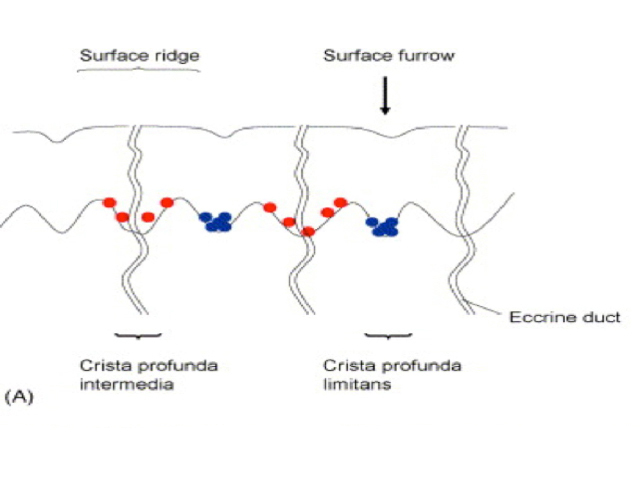
Parallel lines are seen on dermatoscopy on acral skin and reflect the unique anatomy of the dermoepidermal junction in palmar and plantar skin. Surface ridges (on which eccrine ducts open to the surface) overlie the crista profunda intermedia whilst surface furrows overlie the crista profunda limitans (Figure 23). The different sites of predilection for proliferation of acral naevus cells and acral melanoma cells result in parallel furrow and parallel ridge patterns respectively (Figures 24, 25, 27 and 28). Exceptions do, however, occur (Figure 26a and 26b). Acral melanoma may also show structureless pigmentation reflecting proliferation of pigmented melanocytes unrelated to the normal epidermal architecture.

Acral volar skin. Acral naevus

  • Figure 23. Diagram. Preferential proliferation patterns of early melanoma cells and nevus cells in acral volar skin. Early melanoma cells (red dots) are mainly found as individual cells in the crista profunda intermedia under the surface ridge. In contrast, nests of acral nevus cells (blue dots) are seen in the crista profunda limitans under the surface furrow. Takata M and Saida T. J Dermatol Sci 2006; 43: 1-10, with permission
  • Figure 24. Dermatoscopy of acral naevus. Parallel furrow pattern is typical.
  • Figure 25. Histology of acral naevus. Columns of melanin pigment appear within narrow furrows in the skin surface, giving the parallel furrow pattern on dermatoscopy that is characteristic of acral naevi.

Acral melanocytic naevi

  • Figure 26a. Dermatoscopy of lesion reported to be acral naevus. Parallel ridge pattern is unusual in acquired naevi. Unstable acquired melanocytic lesions with parallel ridge pattern should undergo diagnostic excision, as this pattern is more typical of melanoma. The nests of naevus cells are typically present within the crista profunda intermedia. Courtesy Dr Tim Hackett.
  • Figure 26b. Dermatoscopy of acral naevus that also has parallel ridge pattern. The nests of naevus cells are typically present within the crista profunda intermedia. Courtesy Dr Jean Yves Gourhant.

Acral melanoma

  • Figure 27. Dermatoscopy of acral melanoma. Disorder and parallel ridge pattern as seen here or structureless pattern are typical of acral melanoma. Courtesy Dr Alan Cameron.
  • Figure 28. Histology of acral melanoma showing proliferation of single melanocytes in and around the eccrine duct indicating involvement of the crista profunda intermedia. This preferential localization accounts for the typical parallel ridge pattern seen on dermatoscopy.

Nail matrix melanocytic lesions

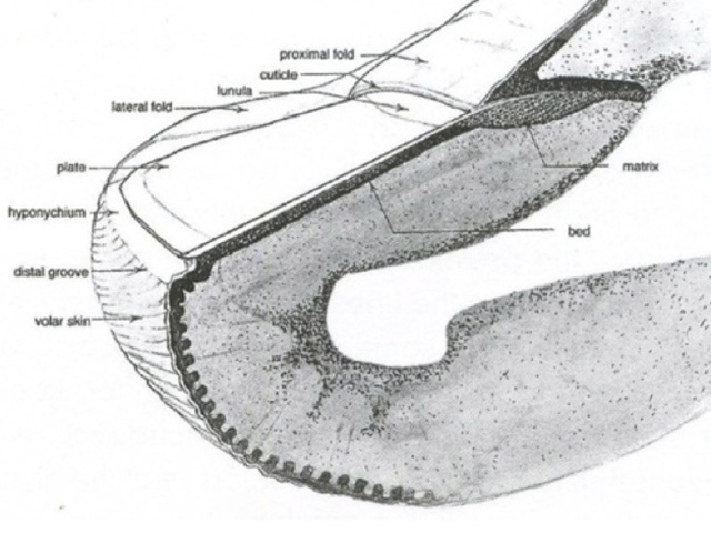
Pigmented nail matrix lesions, including nail matrix melanoma, typically present with longitudinal melanonychia: a pigmented stripe running from the cuticle to the free edge of the nail. The primary lesion arises in the germinal matrix deep to the proximal nail fold and proximal nail plate and melanin is transferred to the developing nail plate and growth of the lesion may manifest as increasing width of the stripe. Extension of the primary lesion to the proximal nail fold (Hutchinson sign) is a clinical clue, albeit relatively late, to the diagnosis of nail matrix melanoma (Figures 29 and 30).

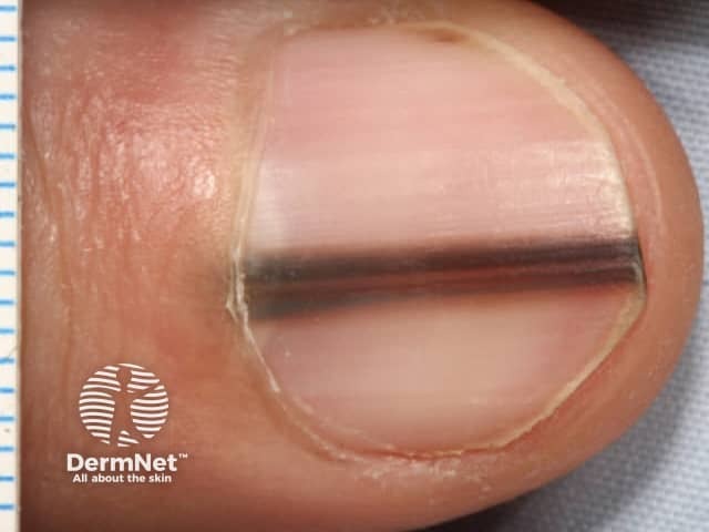
Examination of the pigmented stripe can reveal the hallmark clues of pigmented nail-matrix melanoma: longitudinal melanonychia with lines parallel chaotic; lines parallel varying in width, interval and colour.

Nail matrix melanoma

  • Figure 29. Diagrammatic representation of the nail apparatus. From Kittler et al (1) .
  • Figure 30. Clinical image of a pigmented stripe on the thumb of a 25 year old man. It can be noticed that the base of the pigment stripe is 3mm wide (see scale left extremity of image) while the distal end is 2mm wide. This relates to the size of the tumour at the time each part of the nail was formed. Courtesy Cliff Rosendahl.
  • Figure 31. Dermatoscopic image of the nail shown in Figure 30. Lines parallel vary in width, interval and colour. Courtesy Cliff Rosendahl.
  • Figure 32. Histology of melanoma in situ of nail plate. Rete ridges in the nail matrix vary in width, spacing and pigment density corresponding to the dermatoscopic findings of lines parallel varying in width, interval and colour in the nail plate. Low power view, stained with S100 to demonstrate melanocytes in the nail matrix of the thumb (Figures 30 and 31).

It is essential that nail matrix biopsy be performed competently so that adequate tissue is obtained to facilitate histological diagnosis. One method is the ‘fenestration’ technique (4) but the authors now prefer the ‘pop the bonnet’ technique as this allows direct visualisation of the entire matrix including dermatoscopy of nail bed and nail plate (figure 33a, b).

Nail bed and nail plate dermatoscopy

  • In-vivo dermatoscopy of nail bed (figure 33a) and nail plate (figure 33b) seen during nail matrix biopsy. Images courtesy Dr Luc Thomas.

The biopsy should ideally include matrix from the proximal edge of pigment on the nail matrix to the distal edge of the matrix (located at the lunula). It is essential that nail matrix tissue is examined by a dermatopathologist cognisant of the challenges this can present. A recent publication asserts that immunoperoxidase stains may be necessary to safely exclude malignancy (5).

Patterns of dots

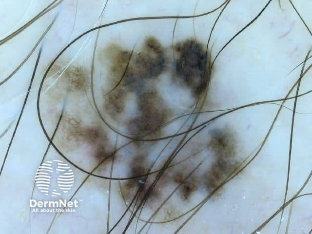
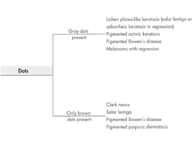
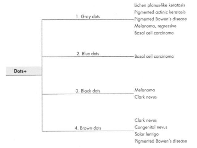
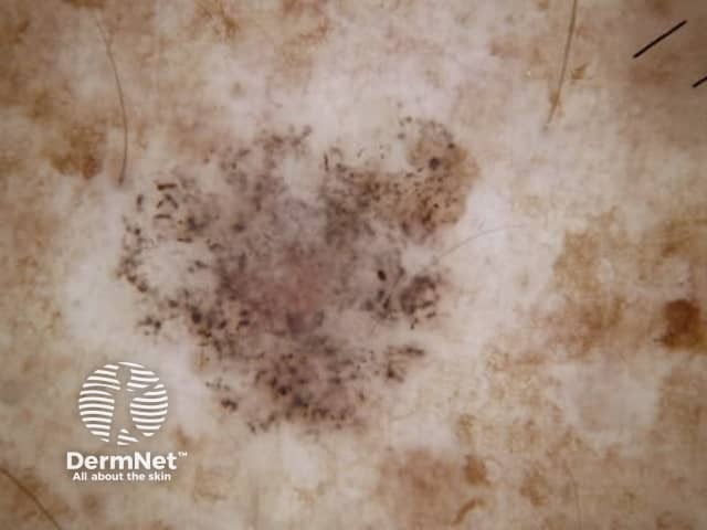
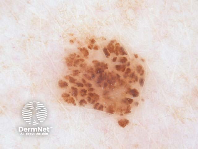
Dots are small enough to have no discernable shape at the magnification provided by a dermatoscope. They represent melanin, or occasionally haemosiderin, with the colour depending on the level of the pigment within the skin in the case of melanin. Dots may be black (superficial epidermal melanin), brown (basal epidermal melanin or superficial dermal haemosiderin), grey (papillary dermal melanin) or blue (reticular dermal melanin). They are usually present in association with another pattern. Brown or grey dots are occasionally found without another pattern (Figures 34-36). Red dots, representing a vascular pattern, are discussed later.

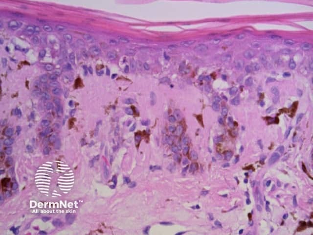
Black dots result either from pagetoid pigmented melanocytes or free pigment within the stratum corneum. Brown dots reflect either small nests of melanocytes in the basal epidermis, focal pigmented keratinocytic proliferation, as seen in some forearm solar lentigines, or superficial dermal haemosiderin deposition. Grey dots are due to melanin pigment within the papillary dermis, either free, in small nests of melanocytes or, more commonly, in melanophages. Blue dots reprresent deeper dermal melanin. The differential diagnosis of dots on dermatoscopy is shown in Figure 34.

Patterns of dots on dermatoscopy

  • Figure 34a,b. Causes of patterns of dots on dermatoscopy. From Kittler et al (1).
  • Figure 35. Dermatoscopy of lichen planus-like keratosis with grey dots centrally. Courtesy Dr Cliff Rosendahl.
  • Figure 36. Histology of lichen planus-like keratosis. The papillary dermal melanophages give rise to the grey dots seen dermatoscopically.

Patterns of clods

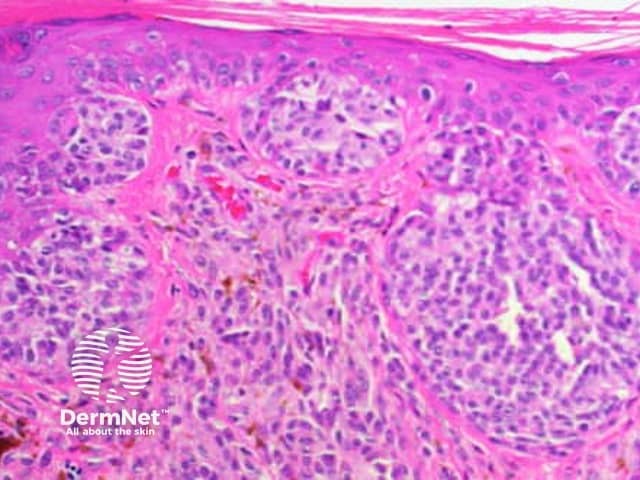
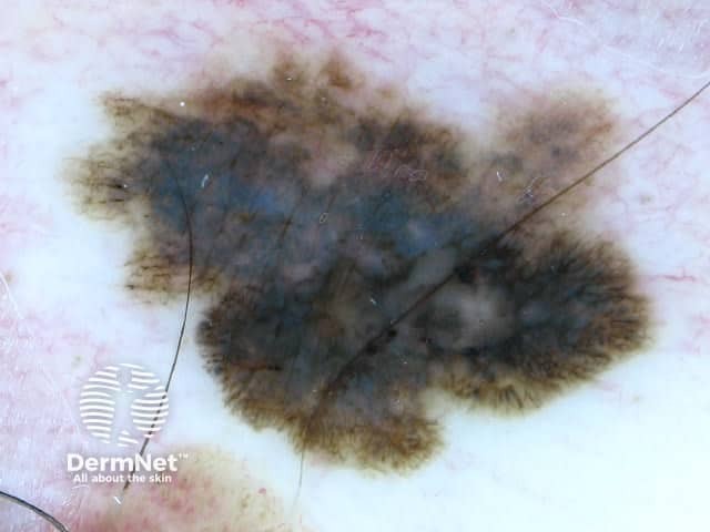
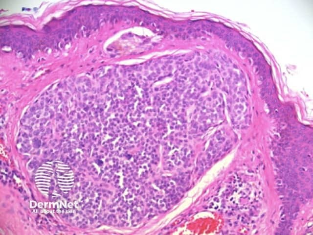
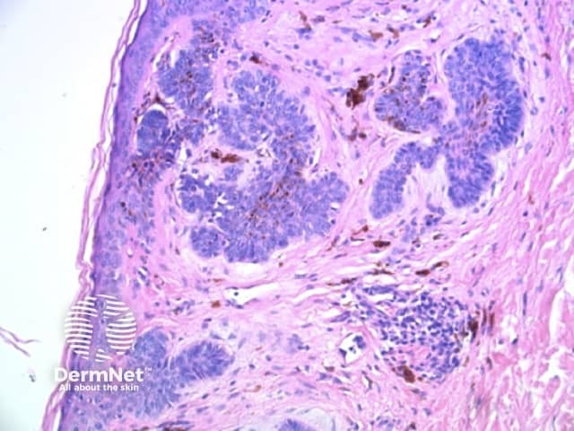
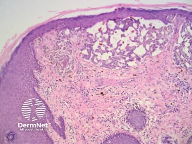
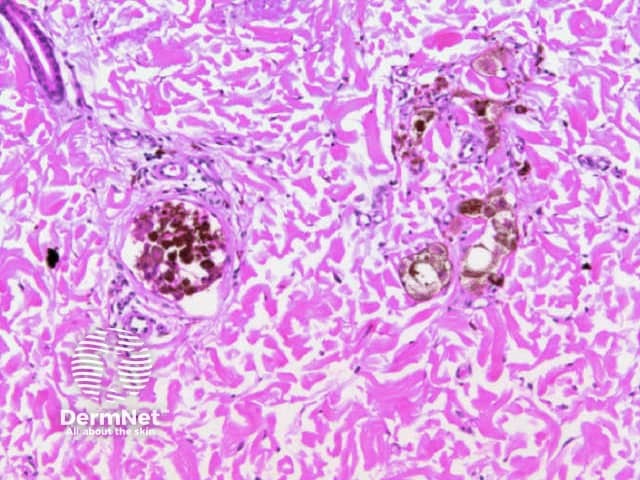
Clods are solid rounded structures of sufficient size to show an appreciable variability in size. Their colour depends on the nature and, in the case of melanin, depth of the substance they comprise (Figures 37-50). Other terms have been used for clods in specific settings such as blue-grey ovoid nests for grey and / or blue clods seen in basal cell carcinoma.

Patterns of clods on dermatoscopy

  • Figure 37a,b. Algorithm for dermatoscopic assessment of clods. From Kittler et al (1).

Red, blue and purple clods (vascular lesions)

  • Figure 38. Dermatoscopy of two adjacent haemangiomas. Clods vary in colour depending on the oxygenation of blood within them. Courtesy Dr Alan Cameron.
  • Figure 39. Histopathology. Red and purple clods in haemangiomas and angiokeratomas are due to the presence of variably oxygenated blood within grouped or ectatic blood vessels.
  • Figure 40. Dermatoscopy of pyogenic granuloma. Red clods are typically separated by white lines. Courtesy Dr Cliff Rosendahl.
  • Figure 41. Dermatoscopy of pyogenic granuloma. The vascular lobules are separated by fibrous tissue corresponding to the white lines on dermatoscopy.

Orange clods (keratin)

  • Figure 42. Histopathology of basal cell carcinoma. Orange clods may represent serum overlying an area of erosion.
  • Figure 43. Dermatoscopy of seborrhoeic keratosis. Orange clods may also be due to keratin containing melanin. Courtesy Dr Cliff Rosendahl.
  • Figure 44. Histopathology of seborrhoeic keratosis. Keratin admixed with variable amounts of melanin pigment in crypts and cysts within the acanthotic epithelium corresponds to the orange, yellow or white clods seen on dermatoscopy.

White, orange to black clods (keratin)

  • Figure 45a. Dermoscopy of dermal naevus. White, yellow and brownish clods due to keratin within dilated follicular infundibula or between surface papillations.
  • Figure 45b. Histopathology of melanocytic naevus. Keratin within dilated follicular infundibula or between surface papillations can give similar appearing clods to those seen in seborrhoeic keratosis.
  • Figure 46a. Dermoscopy of papillomatous (Unna) melanocytic naevus. The skin coloured melanocytic masses form large "cobblestone" skin coloured clods. Keratin between surface papillations appear as yellow to black clods.
  • Figure 46b. Histopathology of Unna naevus. Accumulation of non-pigmented melanocytes in the superficial dermis can eventually lead to papillations, which are separated by keratin-filled epidermal invaginations. These accumulations of melanocytes and papillations give rise to the skin coloured clods typical of melanocytic naevi. Intervening orange, yellow or white clods seen on dermatoscopy are due to keratin in epidermal invaginations between the papillations. Skin coloured clods due to non-pigmented acanthotic keratinocytic proliferation are also seen in some seborrhoeic keratoses.

White clods (sebaceous hyperplasia)

  • Figure 47. Dermatoscopy of sebaceous hyperplasia. White clods are due to large sebacecous glands. Courtesy Dr Cliff Rosendahl.

Colour and depth of clods

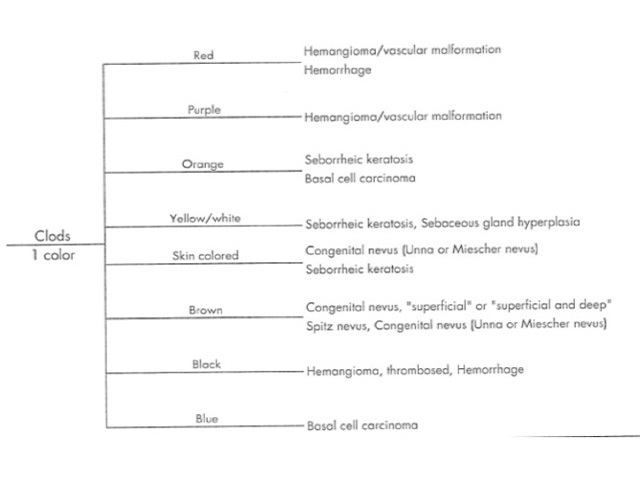
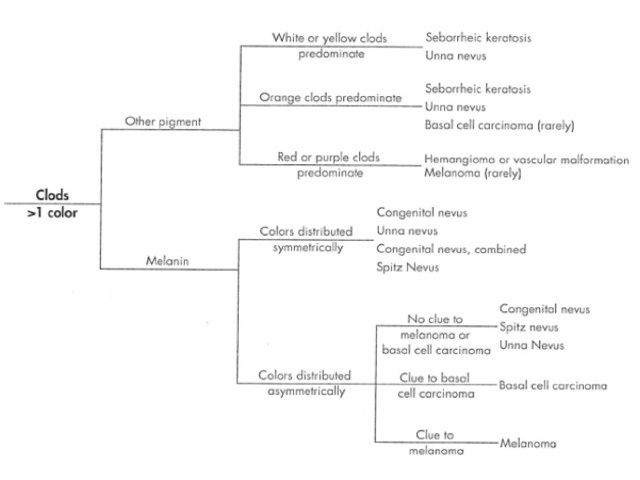
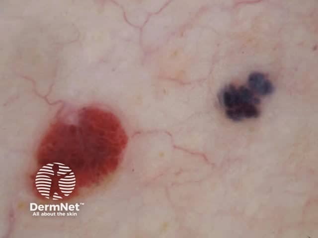
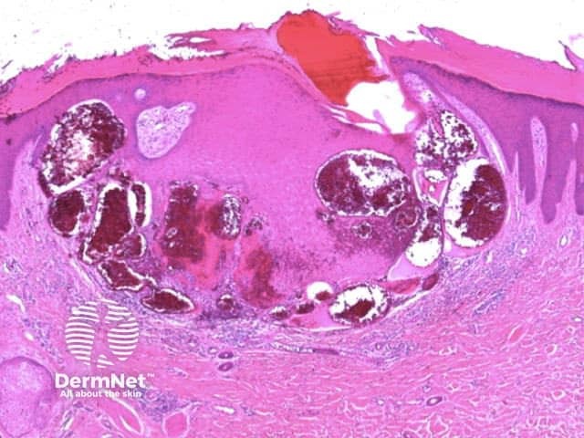
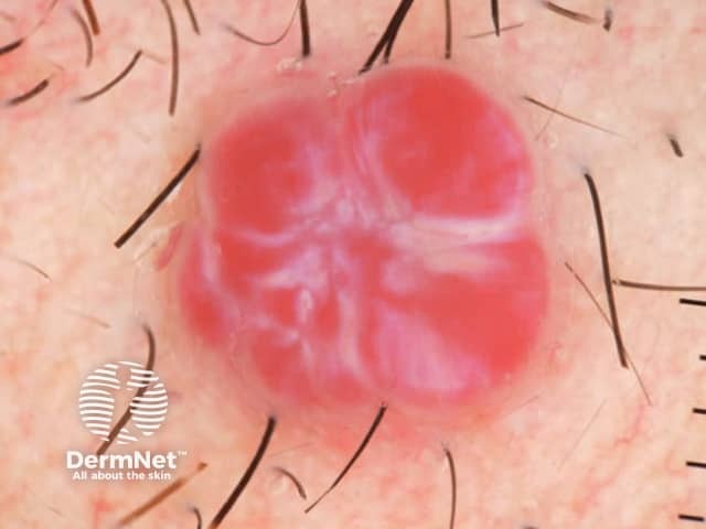
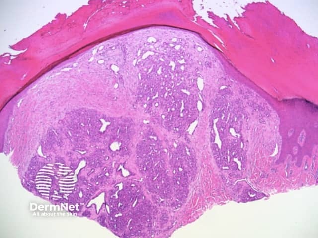
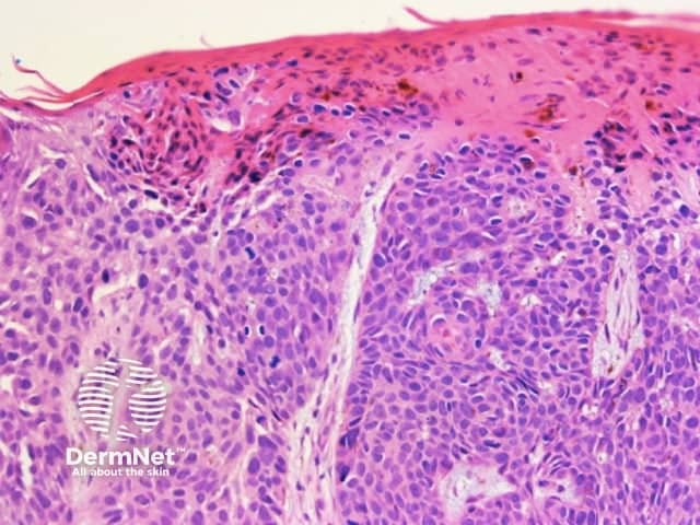
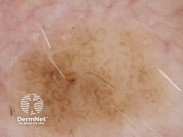
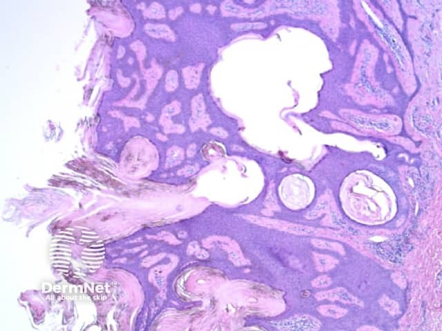
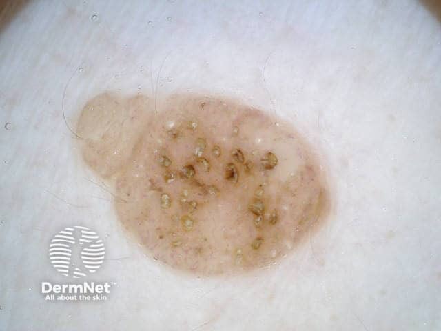
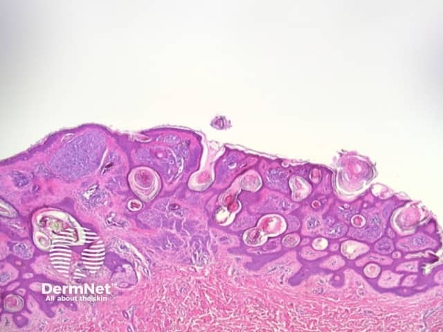
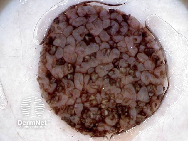
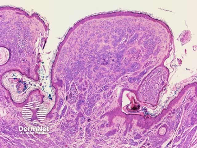
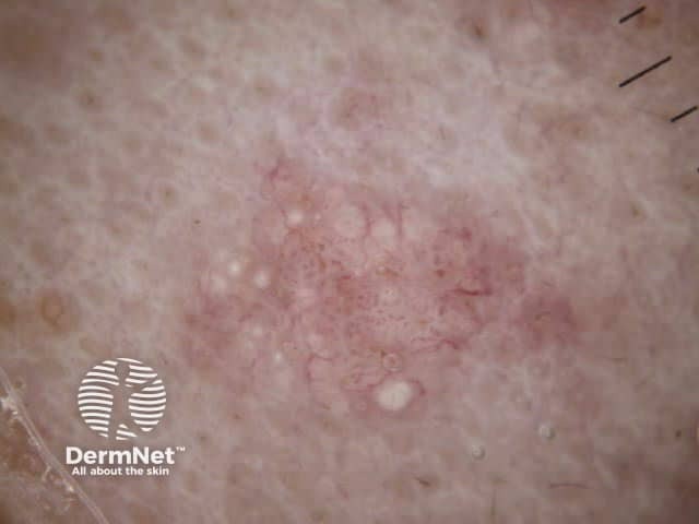
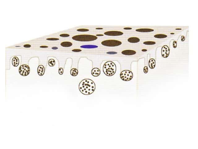
Figure 48. Diagram. Brown, grey and blue clods are due to nests of melanin-containing cells at different levels in the skin. From Kittler et al (1).

Brown clods (melanocytic naevi)

  • Figure 49a. Dermoscopy of dermal naevus showing tightly packed brown clods due to grouped melanocytes under effaced epidermis.
  • Figure 49b. Histopathology of melanocytic naevus (not 49a). Accumulation of non-pigmented melanocytes in the superficial dermis may lead to flattening of the epidermal rete.
  • Figure 49c. Dermoscopy of dermal naevus showing separated brown clods due to dermal nests of melanocytes.
  • Figure 49d. Histopathology of melanocytic naevus (not 49c). Large nests of pigmented melanocytes at the dermoepidermal junction or in the superficial dermis produce brown or grey clods on dermatoscopy.

Blue clods in basal cell carcinoma (deep melanin)

  • Figure 50b. Dermoscopy of basal cell carcinoma. Irregular blue clods are common, as well as branched red lines (blood vessels) and ulceration.
  • Figure 50b. Histopathology of basal cell carcinoma. Blue clods are common and are a clue to the diagnosis. They reflect pigmented nests of basal cell carcinoma in the reticular dermis.

Patterns of circles

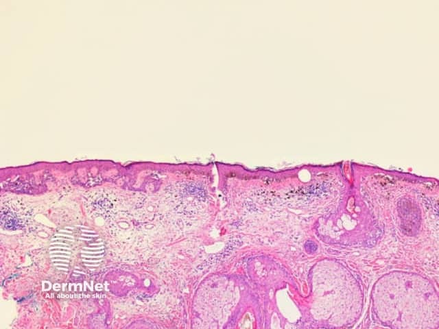
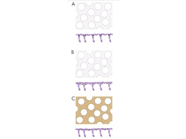
Circles can be seen on non-facial skin where they arise by expansion of rete so that the relatively less pigmented base becomes discernible (Figure 51).

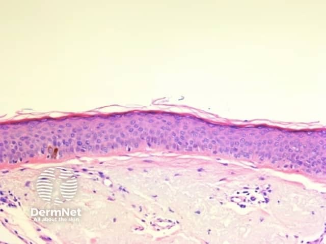
Circles are much more commonly seen on facial skin, reflecting epidermis with flattened rete (Figure 52) with regularly placed pigmented follicular infundibula (Figures 53-59). Differentiation between solar lentigo, pigmented actinic keratosis and melanoma-in-situ on facial skin can be difficult or, in some cases, impossible by dermatoscopy.

Circles in normal skin

  • Figure 51. Diagram. The relationship of non-facial circles to reticular lines is shown. Circles become visible when the epidermal rete are sufficiently wide to permit the less pigmented base of the rete to be visualized. From Kittler et al [1].
  • Figure 52. Histopathology of facial skin. Epidermal rete are typically absent so that the epidermis is featureless from a dermatoscopic perspective and the main feature is follicular openings.
  • Figure 53. Diagram. Circles on facial skin reflect prominent hair follicles with associated pigmentation in an otherwise featureless epidermis. Pigmentation may be within the epithelium giving grey circles (A), in melanophages around hair follicles giving circles comprising grey dots (B) and may also involve the intervening epidermis giving grey circles in a structureless brown background (C). It should be noted that non-pigmented follicular openings do not constitute a pattern of circles. From Kittler et al [1].

Circles in melanoma in situ

  • Figure 54. Dermatoscopy of melanoma-in-situ (lentigo maligna) on facial skin. Grey circles in a featureless brown background. Courtesy Dr Cliff Rosendahl.
  • Figure 55. Histology of melanoma in situ. See Figure 54. The grey circles correspond to pigmented melanocytes within follicular infundibula.
  • Figure 56. Histopathology of melanoma in situ. When perifollicular melanophages are prominent, as in this case, circles may appear to comprise grey dots.

Circles in benign skin lesions

  • Figure 57. Dermatoscopy of facial solar lentigo. These typically have brown and sometimes grey circles. Courtesy Dr Cliff Rosendahl.
  • Figure 58. Histopathology of solar lentigo. Involvement of follicular infundibula manifest as circles on dermatoscopy.
  • Figure 59. Histopathology of pigmented actinic keratosis. Involvement of follicular infundibula may also produce circles on dermatoscopy.

Pseudopods

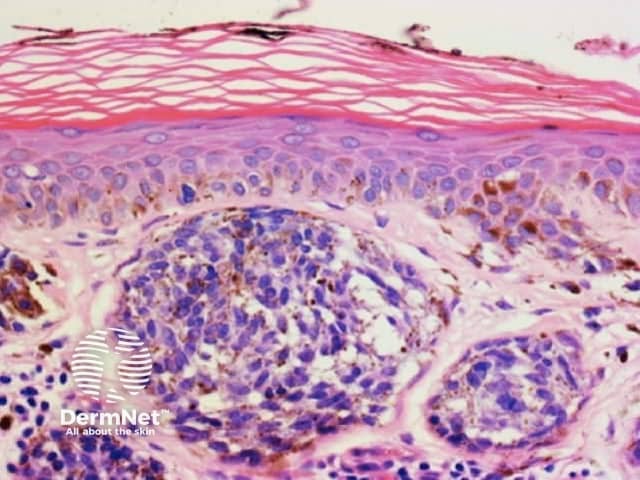
Pseudopods are structures that resemble radial lines but that have a bulbous outer end. They only occur in combination with other patterns with the typical arrangement being pseudopods at the periphery and the other pattern centrally. The second pattern is most often structureless, with clods next in frequency, and reticular lines least common.

Regularly distributed circumferential pseudopods suggests a pigmented spindle cell (Reed) naevus whereas segmental pseudopods create asymmetry and are a clue to melanoma. In both cases, the histologic correlate is large nests of pigmented melanocytes at the periphery of the lesion (Figure 60).

Pseudopods

  • Figure 60a. Diagram. Pseudopods comprise large nests of melaocytes at the periphery of a lesion. From Kittler et al (1).
  • Figure 60b. Histopathology of melanoma. Large peripheral nests of pigmented melanocytes. In this context the pseudopods are segmental creating asymmetry and are a clue to the diagnosis of melanoma on dermatoscopy.

Structureless areas

Structureless areas (Figures 61-64) are seen when:

  1. Pigment has no relationship to the normal epidermal architecture (eg blue naevus, dermal metastatic melanoma, haemorrhage, including most intracorneal haemorrhage; and;
  2. When the epidermis is effaced (see structureless with lines) or rete are absent (see facial skin).

Structureless areas on dermatoscopy

  • Figure 61. Diagram. Causes of structureless areas of a single colour. From Kittler et al [1].

Structureless areas

  • Figure 62. Dermatoscopy of blue naevus. Structureless blue appearance. Courtesy Dr Iris Zalaudek
  • Figure 63. Histopathology of blue naevus. The heavily pigmented dendritic melanocytes are randomly arranged with no relationship to the architecture of the overlying epidermis and so appear structureless on dermatoscopy.
  • Figure 64. Histopathology of pigmented dermal metastatic melanoma. This also appears strucureless and blue, due to the depth of the pigment and lack of relationship to the epidermal architecture.

Clues to diagnosis in dermatoscopy

Using the algorithmic method for dermatoscopic diagnosis of pigmented lesions involves the analysis of pattern and colour to arrive at a differential diagnosis. Clues are then used to favour one diagnosis over another. A clue may be a pattern (eg eccentric structureless zone as a clue to melanoma), an arrangement of pattern / colour (eg central white structureless and peripheral reticular lines or circles in dermatofibroma), a feature too localized to form a pattern (eg blue clods as a clue to pigmented basal cell carcinoma), the absence of a feature, or a particular vascular pattern (eg branched vessels as a clue to basal cell carcinoma).

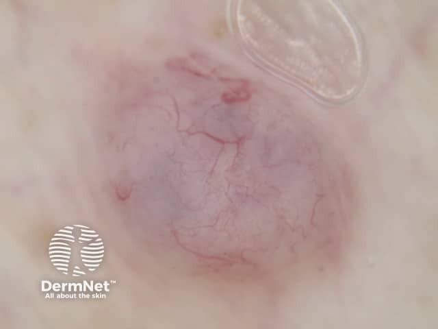
The correlation of many of these clues with histology has been covered in the earlier discussion. The significance of vascular patterns will be addressed in the assessment of non-pigmented lesions. It is important to note, however, that other than in the context of primary vascular lesions presenting as red-purple clods, as discussed previously, there is a poor correlation between histology and vascular patterns on dermatoscopy as these patterns are best appreciated when viewed in three dimensions and from above.

Dermatoscopic histologic correlation of non-pigmented lesions

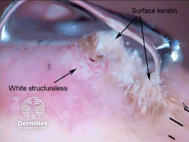
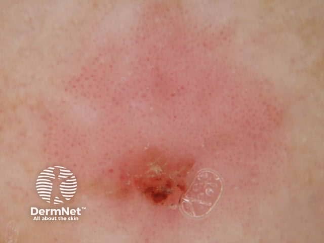
Pigment, when present, is typically used to generate a dermatoscopic differential diagnosis using the algorithms mentioned previously and covered in the DermNet section on dermatoscopic diagnosis. When pigment is not present, other features must be used for diagnosis. The key features used include the presence or absence of non-traumatic ulceration, the recently described “white clues” and vascular patterns (Figures 65-80).

Non-pigmented lesions on dermatoscopy

  • Figure 65. Diagram. The key features used in the dermatoscopic diagnosis of non-pigmented lesions include ulceration, “white clues”, the contour of the lesion and the nature of the blood vessels. Courtesy Dr Cliff Rosendahl.[7] [

White clues

These recently described clues are of great importance in the diagnosis of certain non-pigmented lesions. The term “white clues” refers to white lines or, in a raised lesion (defined as a visibly or palpably raised lesion or a lesion with looped vessels on dermatoscopy), keratin, white circles or white structureless zones. See Figure 72[6].

Abstract of publication describing white areas in SCC

  • Figure 66. Abstract of the paper describing the importance of keratin (assessed clinically) and white circles and white structureless zones (both assessed dermatoscopically) in the dermatoscopic diagnosis of keratoacanthoma and squamous cell carcinoma.[4]

White areas in SCC

  • Figure 67. Dermatoscopy of a squamous cell carcinoma. Keratin and white structureless areas in a raised lesion are clues to the diagnosis of squamous cell carcinoma and keratoacanthoma. Courtesy Dr Cliff Rosendahl.
  • Figures 68a, 68b, 68c. Histopathology of well differentiated squamous cell carcinoma, the same lesion as figure 67. White structureless zones correspond to highly keratinised squamous carcinoma cells massed under the stratum corneum. 68b shows highly keratinised squamous carcinoma cells massed under the stratum corneum and 68c shows surface keratin (hyperparakeratosis). Courtesy Dr Cliff Rosendahl.
  • Figure 69. Dermatoscopy of squamous cell carcinoma. White circles are a further clue to the diagnosis of squamous cell carcinoma and keratoacanthoma. Hairs may be seen eminating from the centre of the circles showing their follicular origin. Courtesy Dr Cliff Rosendahl.
  • Figures 70a, 70b, 70c and 70d show histopathology of the same lesion as figure 69. The histology of 70a shows atrophic folliculosebaceous structures underlying the invaginations (identifiable beneath the far right keratin plug). The white circles seen on dermatoscopy are due to acanthosis and hypergranulosis associated with highly keratinised (well differentiated) squamous carcinoma cells invading hair follicles. Courtesy Dr Cliff Rosendahl.

Vessels as clues

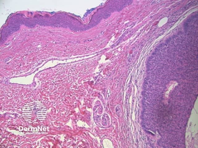
The pattern of blood vessels can be a clue to the diagnosis of pigmented lesions and is a key component of the assessment of non-pigmented lesions by dermatoscopy. In general there is a relatively poor correlation between the dermatoscopic vascular patterns and the histology of particular lesions. This is of little importance as non-pigmented lesions are obviously visible following staining of tissue sections for histology precluding the need to assess secondary features.

Vascular patterns in dermatoscopy

  • Figure 71. Diagram. Vessel types and arrangements as seen dermatoscopically.
    Left – vessel types/ A: Dots, B: Clods, C: Straight, D: Looped, E: Curved, F: Serpentine, G: Helical, H: Coiled.
    Right – vessel arrangements: A: Random, B: Clustered, C: Serpiginous, D: Linear, E: Centred, F: Radial, G: Reticular, H: Branched. From Kittler et al [1].
  • Figure 72. Diagram. There is a relationship between epidermal acanthosis and the pattern of blood vessels observed on dermatoscopy. When the epidermis is of relatively normal thickness dilated papillary dermal blood vessels appear as red dots dermatoscopically. This can be seen in both inflammatory conditions (such as psoriasis) and in neoplasms such as flat seborrhoeic keratosis and intraepidermal carcinoma. As the epidermis becomes progressively more acanthotic these vessels become drawn out to form curved and looped vessels. For this reason the dermatoscopic finding of looped vessels is able to be used in the algorithm for diagnosis of non-pigmented lesions to indicate that a lesion is raised. From Kittler et al [1].

Vascular patterns in intraepithelial carcinoma

  • Figure 73. Dermatoscopy of intraepidermal carcinoma. Vessels may appear as dots or be coiled. Courtesy Dr Cliff Rosendahl.
  • Figure 74. Dermatoscopy of pigmented intraepidermal carcinoma. The same vascular pattern as in Figure 73, seen in conjunction with lines of pigmented dots, arranged radially at the periphery, with structureless brown or grey zones, is typical. Courtesy Dr Peter Bourne.
  • Figure 75. Histopathology of intraepithelial carcinoma. The vessel in the first complete dermal papilla on the left side of the photograph resembles those in psoriasis and would appear as a dot on dermatoscopy.
  • Figure 76. Histopathology of intraepithelial carcinoma. More proliferative superficial dermal vessels manifest as coiled vessels on dermatoscopy.

Vascular patterns in seborrhoeic keratosis

  • Figure 77. Dermatoscopy of acanthotic seborrhoeic keratosis. Looped vessels. Courtesy Dr Cliff Rosendahl.
  • Figure 78. Histopathology of acanthotic seborrhoeic keratosis. Looped vessels are typical of acanthotic lesions.

Vascular patterns in basal cell carcinoma

  • Figure 79. Dermatoscopy of basal cell carcinoma. Branched vessels are a clue. Courtesy Dr Cliff Rosendahl.
  • Figure 80. Histopathology of basal cell carcinoma. One can appreciate the dermatoscopically prominent blood vessels. The branched pattern is not typically seen, as it depends on the chance occurrence of the branch point, the original vessel, and the branches all being in the one plane of section examined.

Activity

Review this article and correlate dermatoscopic and histologic features with each new lesion you excise.


Related information

References

  1. Kittler H, Rosendahl C, Cameron A, Tschandl P. Dermatoscopy. An algorithmic method based on pattern analysis. 2011. Facultas Verlags and Buchhandels AG, Universitatsverlag, Austria.
  2. Ferrara G et al. The influence of clinical information in the histopathologic diagnosis of melanocytic skin neoplasms. PLoS ONE 2009; 4: e5375
  3. Menzies SW et al. An Atlas of Surface Microscopy of Pigmented Skin Lesions: Dermatoscopy (2nd Edition) 2002; p13. McGraw-Hill Book Company, Australia.
  4. Rosendahl C, Cameron A, Wilkinson D, Belt P, Williamson R, Weedon D. Nail matrix melanoma: consecutive cases in a general practice. Dermatol Pract Conc. 2012;2(2):13.
  5. Weedon D, Van Deurse M, Rosendahl C. Occult Melanocytes in Nail Matrix Melanoma. Am J Dermatopathol. In press 2012
  6. Rosendahl C et al Dermatoscopy of squamous cell carcinoma and keratoacanthoma. Arch Dermatol 2012; 148: 1386-92
  7. Rosendahl C, Cameron A, Tschandl P, Bulinska A, Zalaudek I, Kittler H. Prediction without Pigment: a decision algorithm for non-pigmented skin malignancy. Dermatol Pract Concept. 2014;4(1):9

On DermNet NZ

Other websites

Books about skin diseases

See the DermNet NZ bookstore.

Text: Miiskin

Sign up to the newsletter