Main menu
Common skin conditions

NEWS
Join DermNet PRO
Read more
Quick links
Systemic diseases Inflammation
Author: Dr Lucy Webber, Clinical Fellow in Dermatology, Department of Dermatology, University Hospitals Bristol NHS Foundation Trust, UK. DermNet New Zealand Editor in Chief, A/Prof Amanda Oakley, Dermatologist, Hamilton, New Zealand. Copy Editor: Gus Mitchell. September 2017.
Introduction - panniculitis Introduction - panniculitis associated with pancreatic disease Demographics Causes Clinical features Diagnosis Differential diagnoses Treatment Outcome
Panniculitis is inflammation of subcutaneous fat. Despite having diverse causes, most forms of panniculitis have the same clinical appearance.
Pancreatic panniculitis is the inflammation of subcutaneous fat associated with pancreatic disease, most commonly:
Rarely, panniculitis can be associated with other pancreatic disorders, including pancreatic pseudocysts, pancreas divisum and vascular pancreatic fistulas. It has also been reported in patients with human immunodeficiency virus (HIV).

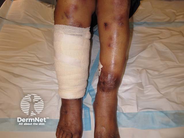
Pancreatic panniculitis

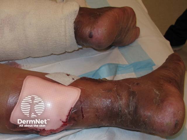
Pancreatic panniculitis

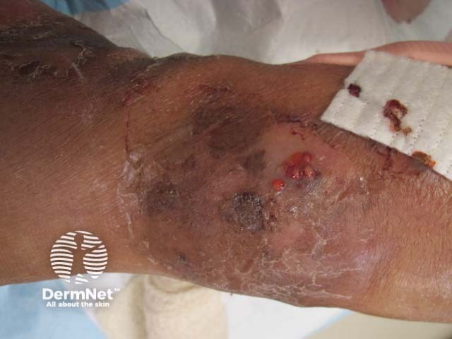
Pancreatic panniculitis
Pancreatic panniculitis is rare and is estimated to occur in 2–3% of all patients with pancreatic disease. Its incidence is higher amongst alcoholic men.
The exact cause of pancreatic panniculitis remains unclear and several mechanisms have been implicated in its pathogenesis. The release of pancreatic enzymes, including lipase, amylase and trypsin, is believed to be the most significant factor.
Other pathogenic factors may include:
Pancreatic panniculitis typically presents with:
Fat necrosis induced by pancreatic enzymes is not always confined to subcutaneous fat and may appear elsewhere, particularly in fat around the joints, which can cause arthritis. The arthritis can affect single or multiple small and large joints and may be migratory, intermittent or persistent, and can progress to necrosis of the cartilage (chondronecrosis) or bones (osteonecrosis).
Panniculitis associated with pancreatic cancer is more likely to ulcerate, persist and recur in comparison to lesions associated with inflammatory pancreatic disease. Cutaneous lesions associated with pancreatic cancer have also been reported to occur in areas other than the lower limbs including the thighs, abdomen, buttocks, arms, scalp and chest.
Panniculitis is diagnosed and classified by a combination of clinical features and skin biopsy findings. In up to 40% of cases of pancreatic panniculitis, skin lesions are the presenting feature of pancreatic disease.
The association of panniculitis with a pancreatic tumour may be recognised by the Schmid triad:
Panniculitis is classified histologically as mostly septal panniculitis (inflammation in the fibrous septa that surround subcutaneous fat lobules) or lobular panniculitis (inflammation of the fat).
Most types of panniculitis have both lobular and septal inflammation. Further classification is based on whether there is subcutaneous vasculitis, and the type of inflammation noted (neutrophils, lymphocytes, histiocytes, or granulomas).
In the very early stages of pancreatic panniculitis, a septal pattern of lymphoplasmacytic inflammation is seen. As the condition progresses, a predominantly lobular panniculitis is seen without associated vasculitis.
The histological hallmark of pancreatic panniculitis is the presence of ‘ghost cells’. These are necrotic adipocytes that turn into an amorphous or granular blue–grey substance. The infiltrate surrounding ghost cells is predominantly neutrophilic, hence pancreatic panniculitis is also termed as neutrophilic panniculitis.
The diagnosis of the underlying pancreatic disease can be made by imaging and blood tests.
Imaging can induce:
The most common form of panniculitis is erythema nodosum. (See panniculitis for the full list of causes and classifications of the disease).
The main clinical differential diagnoses for pancreatic panniculitis are:
The treatment of pancreatic panniculitis should address the underlying pancreatic pathology and may include surgical or endoscopic management. The cutaneous lesions usually heal once this has occurred. In some patients with pancreatic cancer, administration of octreotide (a somatostatin analogue that inhibits pancreatic enzyme production) has resulted in significant improvement of pancreatic panniculitis.
Topical corticosteroids, non–steroidal anti–inflammatory drugs and immunosuppressive drugs are not usually effective treatments for pancreatic panniculitis.
If ulceration occurs, compression hosiery may be required to aid healing.
The outcome for pancreatic panniculitis depends on the underlying disease process. It has a high mortality rate unless the underlying pancreatic abnormality is reversed. The prognosis is particularly poor in cases of pancreatic panniculitis related to pancreatic malignancy.
Several studies have shown the resolution of pancreatic panniculitis after surgical or medical management of the pancreatic disease.
Once the underlying condition is treated, cutaneous lesions will slowly resolve, leaving post–inflammatory changes.