

Stevens Johnson syndrome / toxic epidermal necrolysis: nursing management — extra information
Introduction Demographics Benefits SCORTEN criteria Patient precautions Equipment required Role of health provider Bacteria mapping Intravenous access Pain control Oral feeding
What is Stevens Johnson syndrome / toxic epidermal necrolysis?
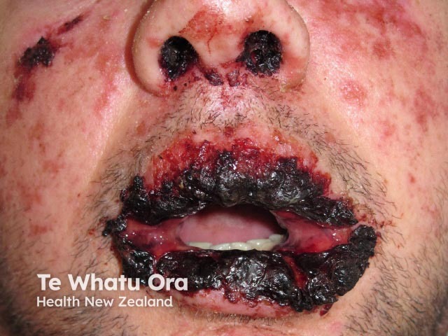
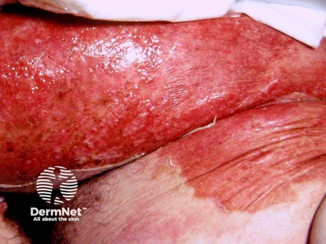
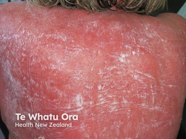
Stevens Johnson syndrome /toxic epidermal necrolysis (SJS/TEN) is a very severe and acute skin disease, almost always caused by a drug.
SJS/TEN is characterised by an extensive necrosis and detachment of the epidermis, which involves skin and mucosal surfaces (genitals, eyes, and mouth).
The nursing care described on this page is adapted from the Créteil protocol for patients with toxic epidermal necrolysis [1].
Who is this protocol for?
This protocol is recommended for all health care providers caring for patients with SJS/TEN or similar skin conditions especially severe cutaneous adverse reactions to drugs.
Suitable nursing care can reduce the mortality and adverse sequelae of SJS/TEN, especially when a large area of skin / mucosa is affected and intensive care is needed. Expert nursing care should commence as soon as the patient has arrived at the hospital and continue until discharge. The patient should be cared for in bed until well enough to ambulate safely.
What are the benefits of this protocol?
The benefits of this protocol are:
- Prevention of bacterial wound infection, which is a very high risk in these patients due to skin impairment
- Prevention of hypothermia
- Improved healing
- Prevention of mucous membrane scarring, avoiding synechiae of eyes and genitals
- Prevention and management of pain.
SCORTEN criteria of severity of SJS/TEN
The prognosis of SJS/TEN should be determined during the first 24 hours. SCORTEN is an illness severity score that has been developed to predict mortality in SJS/TEN. One point is scored for each of seven criteria present at the time of admission. The SCORTEN criteria are:
- Age > 40 years
- Presence of a malignancy
- Heart rate > 120
- Initial percentage of epidermal detachment > 10%
- Serum urea level > 10 mmol/L
- Serum glucose level > 14 mmol/L
- Serum bicarbonate level < 20 mmol/L.
The risk of dying from SJS/TEN depends on the score. Depending on local protocols, if SCORTEN is more than 1, the patient is managed in intensive care, a burns unit or a specialist dermatology unit of a regional hospital.
Estimate total body surface with epidermal detachment
Use the Wallace rule of 9 to estimate the affected body surface area.
Percentages of the total body surface area for an adult or child over 10 years
- Head and neck (front and back): 9%
- Each upper limb (front and back): 9%
- Chest and front of abdomen: 18%
- Back: 18%
- Perineum: 1%
- Each lower limb (front and back): 18%
Percentages of the total body surface area for a child under the age of 1
- Head and neck (front and back): 18%
- Each upper limb (front and back): 9%
- Chest and front of abdomen: 18%
- Back: 18%
- Perineum: 1%
- Each lower limb (front and back): 14%
Over 1 year and below 10 years, the percentage of body surface area changes
- Head decreases by 1% per year
- Lower limbs each increase by 0.5% per year
Precautions in patients with SJS/TEN
Determine any prior history of adverse reaction to antibiotics and antiseptics, as these may be required to manage infection.
Equipment required to manage a patient with SJS/TEN
The SJS/TEN patient should be managed in a single room. Requirements include:
- A bed with air mattress and weighing scale
- A warm cover, eg, emergency warming blanket or space blanket / heat sheet with silver lining
- A thermometer to measure room temperature
- A tympanic or oesophageal thermometer to measure central body temperature
- Hoops to keep the sheets from touching the skin
- Head holder to reduce painful movement
- Wipes to sterilise surfaces
- Disposal bags appropriate for clinical waste.
Personal protective equipment for healthcare providers should include:
- Glasses
- Mask and mob cap
- Gown
- Non-sterile gloves
- Alcoholic solution hand rub, wash or gel.
Patient eye care requirements:
- Sterile ophthalmic compresses
- Vitamin A ointment or other sterile bland eye ointment.
Patient mouth care requirements:
- Small bowl with wadding sticks, tongue depressor
- Bean-shaped bowl to collect spit
- Towel
- One 60 mL syringe with a big tip (if the patient can’t bring a glass to his/her mouth)
- Mouthwash. A suitable prescription is:
- 250 mL bicarbonate1.4%
- 90 mL chlorhexidine 0.12%
- 24 mL nystatin 100 000UI/mL
- 24 mL lidocaine with naphazoline 5%.
Patient genital care requirements:
- Sterile gloves
- Sterile compresses
- Petroleum jelly (yellow soft paraffin or 50% liquid paraffin 50%, white soft paraffin).
Patient skin care requirements:
- Chlorhexidine 0.2% solution
- Sprayer
- Wash cloths
- Hydrocellular dressing
- Paediatric ECG patches
- Petroleum jelly
- Scalpel with #10 blade.
Bacteria mapping requirements:
- Sterile gloves
- Sterile field
- Nine 90-mm agar plates containing 3 different media (white trypticase soy agar for common bacteria; red Columbia agar base with horse blood, nalidixic acid, colistin for gram-positive bacteria; green Drigalski/MAC for gram negative bacteria)
- Sterile kit with 9 sterile ophthalmic compresses + 9 sticks
- Three microbiology request forms (corresponding to 3 skin sites)
- Sterile dressing tray with Kocher forceps.
Intravenous fluid requirements:
- Central catheter kit
- Non-adhesive hydrocellular dressing
- Slim adhesive strips
- Bandages.
What is the role of the health provider?
The health care provider should keep the patient's room clean and warm, and gently but thoroughly cleanse and protect the skin, mouth, eyes and genitals.
Maintain environmental temperature
- The objective is to keep the room temperature 28–32°C, even if the patient has a fever.
- Put a warm cover over the patient, even if the skin/patient feels hot.
- The door should be kept closed.
- The room temperature has to be checked every 6 hours.
- The patient’s central temperature has to be checked every 6 hours with an intra-aural, tympanic or oesophageal thermometer.
Maintain hygiene
- Wash hands with alcoholic solution hand wash before and after care.
- Put on non-sterile gloves before starting each episode of care.
- Ensure correct waste disposal.
Eye care
- Eye care is undertaken 3–6 times each day depending on severity of eye involvement.
- Apply vitamin A or other sterile ocular lubricant ointment generously under the upper and the lower eyelid using one quarter of tube for one eye on each occasion.
- Synechiae can be released by applying the eye ointment.
- Ask the patient to open his/her eyes frequently to avoid synechia formation.
- Do not use saline eye drops or sticks.
- Only use other eye preparations that have been prescribed by an ophthalmologist (including prior prescriptions).
- Put some petroleum jelly on the eyelids if there is crust or erosions
Mouth care
- Mouth care is undertaken 6 times a day.
- Put the mouthwash solution in glass or syringe.
- Ask the patient to gargle with the mouthwash.
- Repeat the gargle three times.
- Spit the solution into the bean-shaped bowl.
- Presoak the stick with the mouthwash solution.
- Use the stick to delicately clean the mucosa of cheek, gum, tongue; change sticks frequently and avoid injury to mucosal lesions.
- Make sure the patient doesn’t swallow the solution.
- Patients can do this care themselves.
Genital care in females
- During the bath
- Clean the genitals delicately with a compress to remove exudate and necrotic mucosa.
- Avoiding injuring the mucosa.
- Sever any synechia between labia minora and labia majora.
- After the bath
- Put a sterile compress into the finger of a sterile glove. Apply petroleum jelly to the outside of the glove.
- The patient or the health provider must put the petroleum-jelly-wrapped compress/glove into the vagina and gently remove it so that the jelly lubricates the lining of the vagina.
Genital care in males
- During the bath
- Pull back the foreskin.
- Clean the genitals delicately with a compress to remove exudate and necrotic mucosa.
- Sever any synechia.
- Avoid injuring the mucosa.
- Replace the foreskin.
- After the bath and 4–6 times a day
- Tap gently with soft wash cloth to dry; do not rub.
- Pull back the foreskin to apply petroleum jelly.
- Replace foreskin.
Skin care rules
There must be more at least two health providers.
- Move the patient using two wash clothes: one maintains the hip the other maintains the shoulder.
- Bacteria mapping has to be done before a bath and is repeated every 48 hours.
- Nitrous oxide inhalation, methoxyflurane and other short-term methods of pain relief can be used to reduce pain.
- The time the patient is naked should be as short as possible.
- Prick blisters every day with a scalpel without remove the top of the blister after the bath.
- Avoid sudden moves and do not scrub the skin.
- Avoid exposing the patient to bright light or TV screen.
Bathing
- Daily bathing should not exceed 15 minutes. It may contain either;
- Antiseptic solution (eg, chlorhexidine 4% for 1.5L of water) if suspicious of infection; or;
- Oatmeal : 3 packets for one bath, if there are dry lesions or crusts.
- Check the water temperature of bath and hand shower.
- Motivate the patient to move by himself to avoid injury during carrying.
- Put the patient under a nursing board.
- Carefully immerse the patient in the bathwater.
- Gently remove dressings, crust, and exudate; avoid scrubbing.
- To clean the skin, tap with a wash cloth rather than rub.
- Rinse with the hand shower, lifting the nursing board above the bath.
- Gently tap to dry using dry wash cloths.
If a bath is contraindicated or unavailable, perform a gentle bed-bath using aqueous cream, warm water and a soft cloth.
During the bath
- Clean mattress with surface wipes and change the sheets.
- If the patient has erosions on his/her back, put a non stick mat onto the bottom sheet (for example hydrocellular dressings joined together by self-adhesive, non-woven fabric sheet).
After the bath
- Lay the patient on the mat.
- Prick, count and record all the blisters.
- Cover the patient with the hoop, sheets, and warm cover.
- Apply petroleum jelly on all parts of the body that are not in contact with hydrocellular dressing, even if skin is not eroded.
Bacteria mapping
Bacteria mapping takes place twice a week BEFORE the patient takes a bath. Two people are needed. Choose three different areas more likely to be infected (eg, axilla, groin, eroded skin). A patient label is applied to each agar plate with date, time, and body site number.
- Open a sterile dressing pack on a tray and lay out 9 ophthalmic compresses and 9 agar plates.
- One operator puts on sterile gloves:
- Holds the tray with one hand;
- Holds a compress with the other hand and applies it to a selected body site;
- Puts the compress onto the first agar plate.
- The other operator:
- Avoids direct contact with the agar;
- Using a wooden stick, presses the compress onto the agar surface for 15 seconds;
- Removes the compress using Kocher forceps;
- Closes the lid of the agar plate immediately.
- The procedure is repeated using the other two agar plates.
- It is then repeated for the other two areas.
- The 9 agar plates are sent immediately to the bacteriology laboratory.
Intravenous access
Insert a central catheter for IV fluids. Follow these precautions:
- Where possible, locate the catheter in an area where skin erosions are minimal.
- Apply hydrocellular dressing to fix the catheter to the patient.
- Avoid adhesive dressing; use 3 thin adhesive strips and a bandage to fix the catheter.
- On removal of a catheter, cut the tip with sterile scissors and send it for bacterial culture.
Fluid requirements
- Must be evaluated during the first 24 hours.
- The usual formula is 2 mL/kg of body weight x percentage of body skin detachment.
Pain control
Evaluate the patient’s pain before, during and after care.
- Inform the patient what to expect and warn him or her that the care may be painful.
- Make the patient comfortable.
- Prescription of analgesics depends of the pain’s intensity and may include paracetamol and opioid. Avoid nonsteroidal analgesics, as these increase risk of skin infection.
- Anxiolytic might also be prescribed if any anxiety is noticed.
Written notes should report every care on the patient record and record pain on a monitoring sheet.
Oral feeding
- Objectives:
- 20–25 kcal/kg daily during the early, catabolic phase
- 25 and 30 kcal/kg daily during the anabolic, recovery phase.
- If the patient doesn't need a nasogastric tube:
- Cool liquid food
- High protein diet.
- If nasogastric tube required:
- Put in the tube very early
- Take great care to minimise injury to affected mucous membranes.
Bibliography
- Protocole de soins des yeux pour les patients ayant une dermatose bulleuse avec une atteinte de la muqueuse oculaire (ou à risque) (syndromes de Lyell et de Stevens- Johnson, érythème polymorphe), Prof Chosidow Olivier, Dr ORO Saskia, Mr Duval Stephane, Mme Colin Audrey.
- Soins aux patients atteints d’un syndrome de Lyell,Mme Rachida Ouedraogo (IDE référente plaies et cicatrisation), Mme Audrey Colin (IDE référente du syndrome de lyell), M. Stéphane Duval (cadre, Service de Dermatologie), Mmes Anne Sophie Rouzière et Chrisitine Geslain (cadres, Service de Réanimation Médicale), Mmes Virginie Devilliers, Amandine Pucci, Emilie Leveque (IDE, Service de Réanimation Médicale).
- Whitney A High, MD et al; Stevens-Johnson syndrome and toxic epidermal necrolysis: Management, prognosis, and long-term sequelae. Updated 19 February 2017. UpToDate.
- de Prost N, Ingen-Housz-Oro S, Duong Ta, Valeyrie-Allanore L, Legrand P, Wolkenstein P, Brochard L, Brun-Buisson C, Roujeau JC. Bacteremia in Stevens-Johnson syndrome and toxic epidermal necrolysis: epidemiology, risk factors, and predictive value of skin cultures. Medicine (Baltimore). 2010 Jan;89(1):28-36. doi: 10.1097/MD.0b013e3181ca4290. PubMed PMID: 20075702. PubMed.
- Shiga S, Cartotto R. What are the fluid requirements in toxic epidermal necrolysis? J Burn Care Res. 2010 Jan-Feb;31(1):100-4. doi:10.1097/BCR.0b013e3181cb8cb8. PubMed PMID: 20061843.
- Creamer D, Walsh SA, Dziewulski P, Exton LS, Lee HY, Dart JK, Setterfield J, Bunker CB, Ardern-Jones MR, Watson KM, Wong GA, Philippidou M, Vercueil A, Martin RV, Williams G, Shah M, Brown D, Williams P, Mohd Mustapa MF, Smith CH. U.K. Guidelines for the management of Stevens-Johnson syndrome/toxic epidermal necrolysis in adults 2016. Br J Dermatol. 2016 Jun;174(6):1194-227. doi: 10.1111/bjd.14530. PubMed PMID: 27317286.
- Duong TA, Valeyrie-Allanore L, Wolkenstein P, Chosidow O. Severe cutaneous adverse reactions to drugs. Lancet. 2017 Oct 28;390(10106):1996-2011. doi: 10.1016/S0140-6736(16)30378-6. Epub 2017 May 2. Review. PubMed PMID: 28476287. PubMed.
On DermNet
- Stevens Johnson syndrome / toxic epidermal necrolysis
- Triggers of SJS/TEN
- Severe cutaneous adverse reaction (SCAR)
- Drug eruptions
- Dermatological emergencies online course
- Bullous drug eruptions
Other websites
- Nécrolyse épidermique - Syndromes de Stevens-Johnson et de Lyell, Protocoles nationaux de diagnostic et de soins — Haute Autorité de Santé, October 2017