

Tuberculids — extra information
Introduction
Demographics
Causes
Clinical features
Complications
Diagnosis
Differential diagnoses
Treatment
Outcome
What is a tuberculid?
A tuberculid is a hypersensitivity reaction presenting as skin changes in association with tuberculosis elsewhere in the body. Mycobacterium tuberculosis organisms cannot be isolated from the tuberculid skin lesions. Four types of tuberculid are usually recognised: erythema induratum (Bazin disease), papulonecrotic tuberculid, lichen scrofulosorum, and nodular tuberculid, and more than one type of tuberculid may be present.
Who gets a tuberculid?
Tuberculids develop in patients with overt or silent tuberculosis infection and an allergy to or a medium-high level of cell-mediated immunity against the tubercle bacillus. They can sometimes appear after initiating anti-tuberculous treatment or after starting antiretroviral treatment for HIV. Tuberculids are mostly seen in countries where tuberculosis remains common, such as China, the Indian subcontinent, and sub-Saharan Africa.
What causes a tuberculid?
Tuberculids are usually associated with Mycobacterium tuberculosis infection, but have also been reported less often following BCG immunisation or in association with atypical mycobacterial infections such as M. avium. It is thought to be due to haematogenous spread of mycobacterial antigens to the skin resulting in a type III or type IV hypersensitivity reaction.
What are the clinical features of tuberculids?
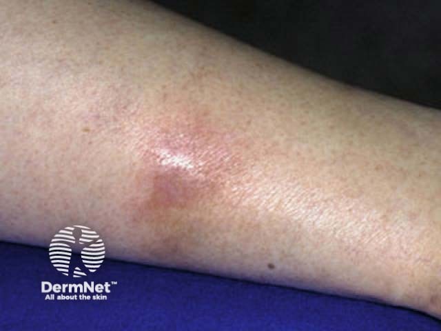
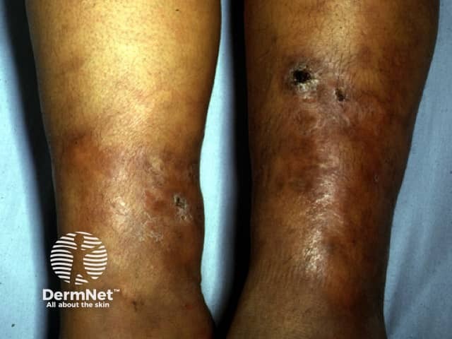
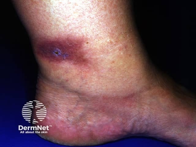
Erythema induratum
Erythema induratum (Bazin disease) is the most commonly recognised form of tuberculid. It typically presents on the backs of the legs of young to middle-aged women as tender or painful indolent blue-red nodules and plaques which may ulcerate and scar.
Papulonecrotic tuberculid
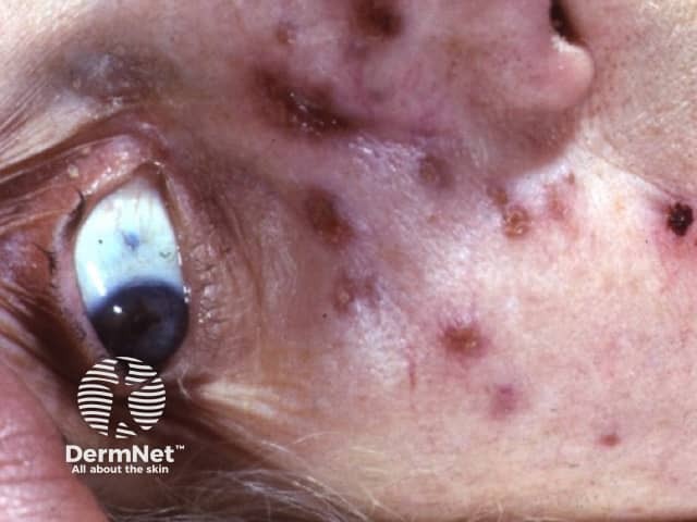
Papulonecrotic tuberculid (tuberculosis papulonecrotica) presents as recurring crops of clustered dusky or erythematous papules and nodules that can become pustular or necrotic to form small ulcers that heal with varioliform (chickenpox-like) scarring after about 6 weeks. The skin lesions develop symmetrically mostly on the limbs of children and young adults before the age of 30 years.
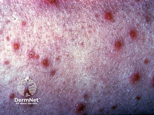
Lichen scrofulosorum
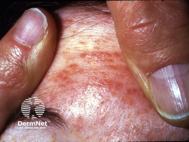
Lichen scrofulosorum (tuberculosis cutis lichenoides) usually affects children and adolescents with tuberculosis of the bone or lymph nodes. It presents as asymptomatic small, slightly scaly, follicular papules on the trunk which can be subtle. The papules are often skin-coloured, but may be yellow or red-brown, and clustered, grouped into an annular arrangement, or coalesced to form plaques, resembling lichen planus or granuloma annulare.
Nodular tuberculid
Nodular tuberculid is probably a variant of erythema induratum presenting as asymptomatic dusky-red papules and nodules.
What are the complications of a tuberculid?
Tuberculids are a complication of tuberculosis. Tuberculid skin lesions can result in:
- Hyperpigmentation
- Scarring
- Chronic ulcer.
Rarely, the eyes may also be involved in a tuberculid.
How is a tuberculid diagnosed?
-
Skin biopsy, including staining for acid-fast bacilli although this is usually negative.
- Erythema induratum: granulomatous lobular panniculitis with neutrophilic vasculitis and caseation necrosis.
- Papulonecrotic tuberculid: lymphohistiocytic small vessel vasculitis with thrombosis, ulceration, and wedge-shaped necrosis.
- Lichen scrofulosorum: noncaseating peri-appendageal granulomas in the superficial dermis.
- Nodular tuberculid: granulomatous inflammation at the junction of the dermis and subcutaneous fat.
- PCR of skin biopsy may demonstrate mycobacterial DNA, particularly in erythema induratum.
- Tissue culture for mycobacteria is negative.
- Positive tuberculin (Mantoux) test — dilute as the reaction can be strongly positive.
- Positive interferon-gamma release assay (IGRA) such as QuantiFERON-TB gold.
- Extensive investigation for underlying tuberculosis, in addition to family history and clinical features; sometimes, the focus of active tuberculosis is cutaneous.
What is the differential diagnosis for tuberculids?
- Erythema induratum: erythema nodosum and other forms of panniculitis
- Papulonecrotic tuberculid: pityriasis lichenoides
- Lichen scrofulosorum: lichen nitidus, lichen spinulosus, sarcoidosis.
What is the treatment for a tuberculid?
Tuberculids characteristically respond to appropriate multi-drug anti-tuberculous treatment.
- Erythema induratum: slow resolution over months with treatment that is only useful where underlying TB has been substantiated. Compression therapy, leg elevation, and NSAIDs may give symptomatic relief.
- Papulonecrotic tuberculid: response begins after 3–4 weeks of treatment.
- Lichen scrofulosorum: rapid response in 2–4 weeks, heals without scarring.
What is the outcome for tuberculids?
Untreated tuberculosis may cause chronic tuberculids with recurrent crops of skin lesions or chronic ulcers. Adequate treatment of the associated tuberculosis infection using appropriate multi-drug anti-tuberculous therapy results in resolution of tuberculids.
Bibliography
- Choi MS, Hong SP, Park BC, Kim MH. Multiple skin colored nodules on both legs in patient with positive QuantiFERON®-TB gold test. Ann Dermatol. 2017;29(1):95–9. doi:10.5021/ad.2017.29.1.95. PubMed Central
- Kajal NC, Prasanth P, Dadra R. Lichen scrofulosorum: an uncommon manifestation of a common disease. Int J Mycobacteriol. 2020;9(3):313–15. doi:10.4103/ijmy.ijmy_41_20. PubMed
- Meghana V, Saravanan G, Karthikeyan K. Papulonecrotic tuberculid. Am J Trop Med Hyg. 2017;97(4):987–8. doi:10.4269/ajtmh.17-0377. Journal
- Patterson JW. Weedon’s Skin Pathology, 5th edn. Elsevier, 2020, pp690–1.
- Santos Felisberto V, Delgado AR, Pinto H, Morais P. Erythema induratum of Bazin: a case of chronic unilateral erythematous plaques in a lower limb. Isr Med Assoc J. 2020;11(22):720–1. Journal
- Singh PY, Sinha P, Baveja S, Sood A. Immune-mediated tuberculous uveitis - a rare association with papulonecrotic tuberculid. Indian J Ophthalmol. 2019;67(7):1207–9. doi:10.4103/ijo.IJO_1550_18. PubMed Central
- Yates VM, Walker SL. Mycobacterial infections. In: Griffiths C, Barker J, Bleiker T, Chalmers R, Creamer D (eds). Rook’s Textbook of Dermatology, 9th edn, 2016: pp27.25–31.