Main menu
Common skin conditions

NEWS
Join DermNet PRO
Read more
Quick links
Last Reviewed: February, 2025
Author: Hon A/Prof Amanda Oakley, Dermatologist, Hamilton, New Zealand, 1997. Updated October 2015. Revised September 2020.
Reviewing dermatologist: Dr Ian Coulson, United Kingdom (2025)
Introduction
Demographics
Causes
Clinical features
Diagnosis
Treatment
Outlook
Granuloma annulare (GA) is a common inflammatory skin condition typified clinically by annular, smooth, discoloured papules and plaques, and necrobiotic granulomas on histology. Granuloma annulare is more correctly known as necrobiotic papulosis.
Granuloma annulare is seen most commonly on the skin of children, teenagers, or young adults. The generalised form is more likely to be found in older adults (mean age 50 years). There is a female predominance of 2:1 over males.
Granuloma annulare may be a delayed hypersensitivity reaction to a component of the dermis or a reaction pattern to numerous triggers. Reported triggering events have included many skin infections and infestations, and types of skin trauma. Inflammation is mediated by tumour necrosis factor alpha (TNFα). The reason this occurs is unknown.
There have also been a number of associations reported with systemic conditions including autoimmune thyroiditis, diabetes mellitus, other organ-specific autoimmune diseases, hyperlipidaemia, and rarely with lymphoma, HIV infection and solid tumours.
Granuloma annulare can occur on any site of the body and is occasionally widespread. It only affects the skin. Granuloma annulare may cause no symptoms, but affected areas are often tender when knocked. The plaques tend to slowly change shape, size, and position.
The localised form is the most common type of granuloma annulare in general, and specifically in children. One or more skin coloured or red bumps form rings in the skin over joints, particularly the knuckles. The surface is smooth and the centre of each ring is often a little depressed. Localised granuloma annulare usually affects the fingers or the backs of both hands, but is also common on top of the foot or ankle, and over one or both elbows. Annular rings may be solitary or multiple, and grow outwards maintaining the ring shape before eventually clearing.

Granuloma annulare
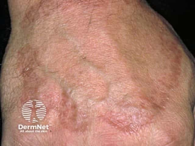
Granuloma annulare
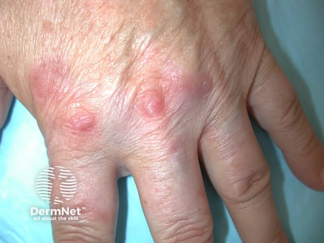
Granuloma annulare
See more images of granuloma annulare ...
Generalised granuloma annulare usually presents in adults, as widespread skin-coloured, pinkish or slightly mauve-coloured patches. The disseminated type is composed of small papules, usually arranged symmetrically in poorly-defined rings 10 cm or more in diameter. They are often found around the skin folds of the trunk (armpits, groin). Itch is common. This is the commonest form associated with HIV.
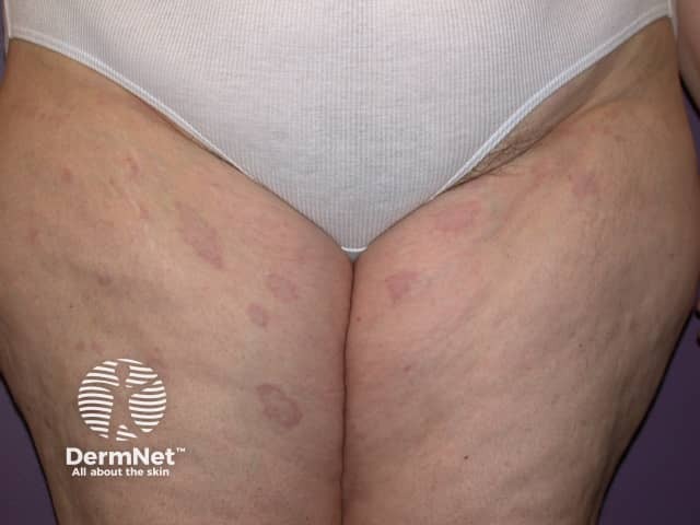
Granuloma annulare
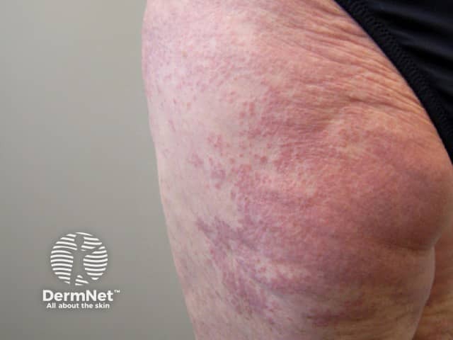
Granuloma annulare
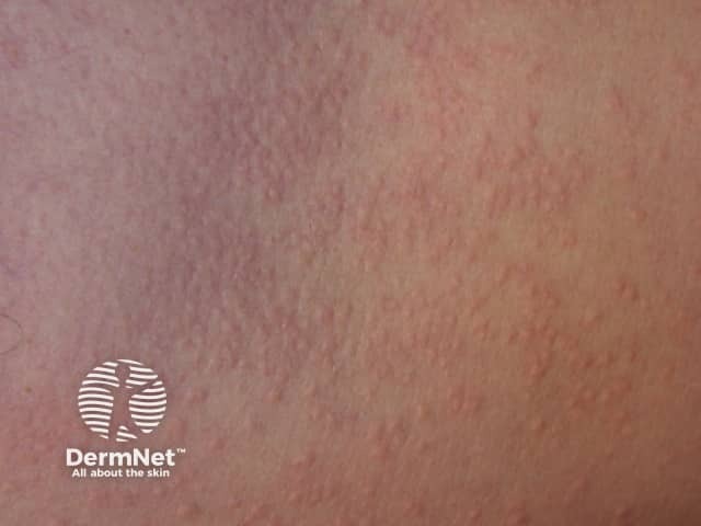
Granuloma annulare
Subcutaneous granuloma annulare is an uncommon form seen mainly in children. It presents as rubbery lumps (nodules) on scalp margins, fingertips, and shins. Subcutaneous granuloma annulare is also called pseudo-rheumatoid nodules because the subcutaneous lesions look rather like rheumatoid nodules; however, they do not arise in association with rheumatoid arthritis.
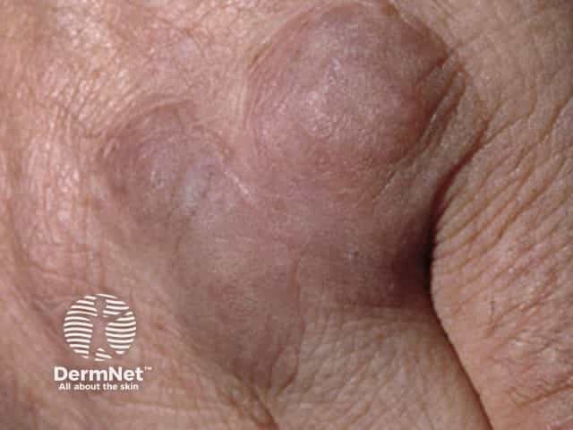
Granuloma annulare
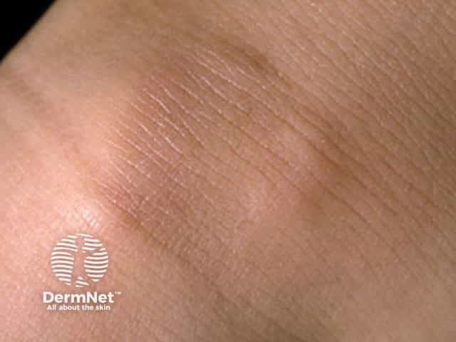
Granuloma annulare
Perforating granuloma annulare presents as yellowish plaques or papules on which a crust forms due to the elimination of damaged collagen through the epidermis. Perforating granuloma annulare is usually localised to the hands, but plaques may occasionally arise on any body site, especially within scars. Dermoscopy helps to confirm the presence of perforations in small papules within otherwise typical plaques of granuloma annulare. Perforating lesions are frequently itchy or tender. Perforating granuloma annulare is uncommon except in ethnic Hawaiians and in association with HIV. All age groups can be affected.
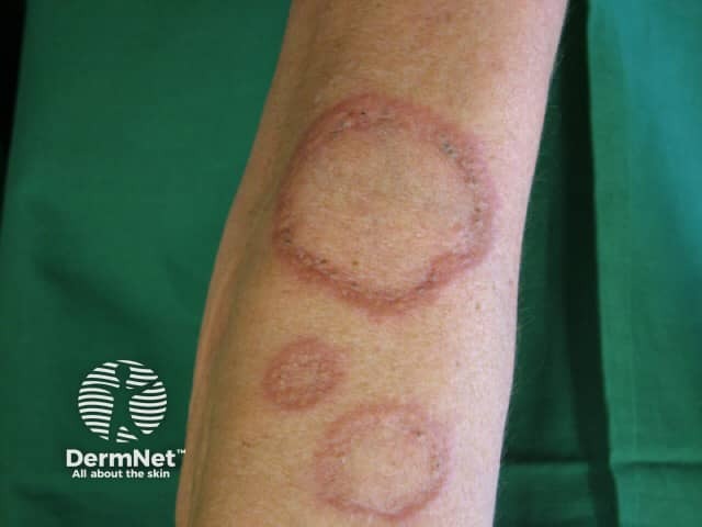
Granuloma annulare
Atypical granuloma annulare describes:
Interstitial granulomatous dermatitis is a finding noted on histology in some patients with extensive granuloma annulare or other disorders with similar clinical presentation.
Granuloma annulare is usually diagnosed clinically because of its characteristic appearance. But sometimes the diagnosis is not obvious, and other conditions may be considered. Skin biopsy usually shows necrobiotic degeneration of dermal collagen surrounded by an inflammatory reaction. See granuloma annulare pathology.
Granuloma annulare is often initially misdiagnosed as tinea because of the annular appearance; the lack of surface scale should lead away from this and other scaly rashes such as discoid eczema or psoriasis. Actinic granuloma is considered by some to be a photo-aggravated variant of granuloma annulare, and by others to be a distinct entity.
In most cases granuloma annulare, does not require treatment because the patches disappear by themselves in a few months, leaving no trace. However, sometimes they persist for years. Treatment is not curative but may help individual lesions.
Options to consider include:
Systemic therapy may be considered in widespread granuloma annulare. The following treatments have been reported to help at least some cases of disseminated granuloma annulare. None of these can be relied upon to clear it, and there are potential adverse effects.
Individual lesions of localised granuloma annulare tend to clear within a few months or years, although they may recur even at the same site. Generalised and atypical variants are more persistent, sometimes lasting decades.