DermNet provides Google Translate, a free machine translation service. Note that this may not provide an exact translation in all languages

Translate

Dermoscopy

The dermoscopy report

Created 2008.

Learning objectives

  • Create a dermoscopy report

Introduction

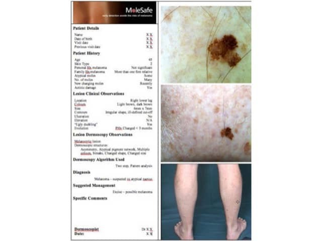
An objective dermoscopy report may be issued for each lesion of concern, and is particularly necessary when the dermoscopy is not performed by the patient's usual primary health practitioner or dermatologist.

A standardised way of reporting dermoscopic findings was described at the 1st World Congress of the International Dermoscopy Society (IDS) in May 2006. A consensus document produced by the Board of the IDS is to be published in the Journal of the American Academy of Dermatology during 2006. It includes ten points, categorised as either recommended or optional.

Lesions of concern

A lesion of concern may be identified by the patient, a family member, a health professional or an expert dermoscopist. Reasons the patient may be concerned about the lesion may include:

  • Cosmetic appearance
  • Symptoms: itch, soreness, proneness to injury
  • Recent trauma
  • Belief that it resembles melanoma
  • Growth of the lesion
  • Apparent change in shape, size or appearance

Clinical concern relating to melanoma may arise because of:

  • Past history of melanoma and/or other risk factors for skin cancer
  • Single or multiple atypical naevi
  • Reliable history of change
  • Irregular shape
  • Irregular colour
  • Large lesion (diameter >6-7mm)
  • Inflammation
  • Ulceration

Alternatively, non-melanoma skin cancer may be present.

Dermoscopic concern may arise because of:

  • Two or three criteria from 3-point checklist: asymmetry, atypical network, blue-white structures
  • Observed growth or change in atypical lesion
  • Specific dermoscopic features of melanoma
  • Specific dermoscopic features of non-melanoma skin cancer

Which melanocytic lesions should be excised?

Lesions with the typical clinical and/or dermoscopic characteristics of melanoma should be excised with a 2-mm margin and the specimen sent for pathology. To guide the pathologist to evaluate a small area that is of concern, orientate the specimen using edge incisions, sutures or ink, and draw a map on the request form. Alternatively, make a superficial round incision using a 1 to 2-mm micropunch in the area of interest and leave the punch in place.

Atypical lesions that do not have diagnostic features for melanoma may also be excised for histology. Melanoma can arise from a naevus, but in about 70% of cases arises de novo. Initially de novo melanoma may lack diagnostic features of melanoma and may also lack features of naevi.

  • Single atypical lesions
  • Nodular atypical lesions
  • Changing atypical lesions

Whole body photographs and digital dermoscopic monitoring may be preferred for:

  • Multiple mildly atypical lesions (12-month intervals)
  • Changing naevus without atypia (3- to 6-month intervals)
  • Moderately atypical lesions (3-month interval)

If no dermoscopic change is noted at 12 months, the lesion can be confidently diagnosed as a naevus.

The International Dermoscopy Society considers that dermatologists should aim to receive histology reports of 5 to 10 benign melanocytic lesions to every melanoma. Less than that should prompt more excisions, and more than 10 benign lesions to every melanoma should prompt more monitoring. However there are inevitably differences in individual practices, and lesions may be removed for reasons that do not directly relate to the risk of malignancy (e.g. cosmetic reasons or to relieve anxiety).

Standardised report

A dermoscopy report should include the following information:

  1. Relevant clinical information about the patient
    E.g. age, history of the lesion, personal and family history of skin cancer and the presence of atypical naevi (recommended).
  2. Clinical description of the lesion
    Location, symmetry, borders, colour, size, elevation (recommended).
  3. The two-step method
    List dermoscopic criteria that differentiate melanocytic from non-melanocytic tumours (I consider this optional).
  4. Dermoscopic description
    Use the standardised terms listed in the Dermoscopy Consensus Report published in 2003, or terminology of modified pattern analysis, and define any new term used.
  5. Algorithm used
    Name which algorithm was used to differente between benign and malignant melanocytic tumours (optional).
  6. Imaging equipment and magnification
    State the brand name and manufacturer of the device (optional).
  7. Images
    Include clinical and dermoscopic views of the tumour (optional).
  8. Diagnosis
    State the diagnosis or differential diagnosis (recommended).
  9. Suggested management
    Management of the lesion may include follow-up, biopsy or excision (recommended).
  10. Comments for the pathologist
    It may be useful to orientate the lesion to guide appropriate sectioning of the specimen when it is excised (optional).
Standardised reports (©MoleMap NZ)

Activity

Compose a dermoscopy report for a lesion of concern.

Page 15 of 18. End of topics. Back to: Dermoscopy contents.

New topic – Melanocytic naevi: new classification


Related information

References

  • Dermoscopy report: proposal for standardization. Results of a consensus meeting of the international dermoscopy society (IDS) Malvehy J, Puig S, Argenziano G, Marghoob AA, Soyer HP. J Am Acad Dermatol. 2006 . Review. Medline.
  • Dermoscopy of pigmented skin lesions: results of a consensus meeting via the Internet. J Am Acad Dermatol. 2003 May;48(5):679-93. Review. Medline.

On DermNet NZ

Other websites

Books about skin diseases

See the DermNet NZ bookstore

Sign up to the newsletter