DermNet provides Google Translate, a free machine translation service. Note that this may not provide an exact translation in all languages

Translate

Whipple disease

Author: Dr Eugene Tan, Dermatology Registrar, Waikato Hospital, Hamilton, New Zealand, 2009.

DermNet NZ Update June 2021


toc-icon

What is Whipple disease?

Whipple disease is a very rare bacterial infection that can affect many organ systems of the body. The disease is named after George Hoyt Whipple who described a case in 1907, but it was not until 1961 that an infection was recognised as the cause.

What is the cause of Whipple disease?

The cause of Whipple disease is the ubiquitous bacterium Tropheryma whipplei (T. whipplei), a gram positive rod. T. whipplei is widespread; it has been detected in soil, sewage water, and faecal material. It is an intracellular bacterium that infects macrophages causing secretion of interleukin (IL)-16, macrophage apoptosis, and loss of mitochondrial membrane potential. The organisms characteristically accumulate in macrophages in the duodenal mucosa. It is spread via the faecal-oral route.

Who gets Whipple disease?

Whipple disease is typically described in Caucasian men with a mean age of 50 years at the time of diagnosis. It is very rare with a global incidence estimated to be 12 cases per year. The prevalence is highest among sewage plant workers, farmers, and agricultural workers, or those with poor hygiene habits such as the homeless. 

In some studies, up to 35% of unaffected healthy individuals carry T. whipplei in their intestines. Not all infected individuals have symptoms. The symptoms of Whipple disease are related to the immune system and its interaction with the bacterium.

Specific defects in the immune system may allow T. whipplei to disseminate widely throughout the body and cause disease.


What are the clinical features of Whipple disease?

The four classic clinical features of Whipple disease are:

  • Arthralgia (joint pain)
  • Weight loss
  • Diarrhoea – due to malabsorption (inability to absorb nutrients through the gastrointestinal tract)
  • Abdominal pain

However, Whipple disease can affect any organ system and has a broad spectrum of clinical features. Chronic localised forms of Whipple disease are being increasingly identified.

Clinical features of Whipple disease
Central Nervous System
  • Eye movement disorders
  • Dementia or memory impairment
  • A decreasing level of consciousness
  • Confusion
  • Insomnia
  • Epilepsy and seizures
  • Imbalance of movement
  • Headache
Musculoskeletal system
  • Arthritis (swollen joints)
  • Arthralgia (painful joints)
Cardiovascular system
  • Endocarditis (inflamed heart or heart valves)
  • Chest pain
Non-specific symptoms
  • Fever
  • Enlarged lymph glands

What are the cutaneous features of Whipple disease?

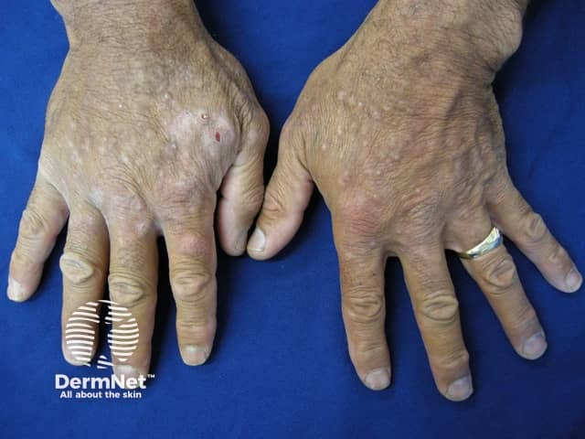
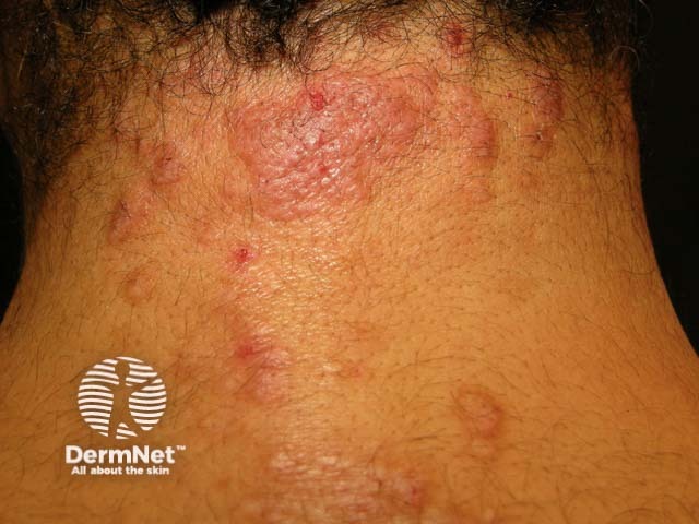
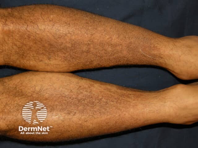
Skin signs in Whipple disease are very rare. They may be due to malnutrition or to an immune reaction to the infection.

Malnutrition-related skin signs are due to malabsorption and diarrhoea.

  • Hyperpigmentation – due to pellagra (niacin/vitamin B3 deficiency), vitamin B12 deficiency or low cortisol levels
  • Petechia or purpura – due to vitamin C or vitamin K deficiency
  • Secondary palmoplantar keratoderma or ichthyosis – due to cholesterol deficiency
  • Oedema – due to loss of protein from the gastrointestinal tract

Inflammatory skin changes are due to the immune response towards T. whipplei. They may resemble:

Skin lesions in Whipple disease

How is Whipple disease diagnosed?

The nonspecific symptoms and signs of Whipple disease together with its rarity make it a very difficult disease to diagnose.

Tissue biopsy is often necessary. The following tests may detect T. whipplei within the tissue:

  • Polymerase chain reaction (PCR) – picks up tiny amounts of bacteria
  • Periodic-Acid Schiff (PAS) – stains the bacteria pink
  • Electron microscopy (EM) – high magnification reveals the bacteria
  • Immunohistochemistry stains – specific antibodies stain T. whipplei.

What is the treatment for Whipple disease?

Infected individuals often need high doses of antibiotics in case the brain is infected by T. whipplei. An induction regime with intravenous ceftriaxone 2 g once daily is given for two weeks.

Endocarditis and CNS involvement require a longer course of ceftriaxone or penicillin G.

Longterm antibiotics such as trimethoprim+sulphamethoxazole should then be continued for up to one year to ensure complete clearance of T. whipplei. Followup upper GIT endoscopy is recommended before ceasing treatment

What is the outcome for Whipple disease?

Before the recognition of a bacterial cause and the use of antibiotics, Whipple disease was invariably fatal.

Treatment can be complicated by a Jarisch-Herxheimer reaction in the first few hours or the immune reconstitution inflammatory syndrome (IRIS) in the first two weeks.

After treatment, a 4-5 year (or longer) period of latency may ensue before recurrence in 10-15% if the infection is not completely eradicated particularly in patients with CNS involvement.


References

  • Canal L, Fuente Dde L, Rodriguez-Moreno J, Penin RM, Marcoval J. Specific cutaneous involvement in Whipple disease. Am J Dermatopathol. 2014;36(4):344–6. doi:10.1097/DAD.0000000000000008. PubMed
  • Crews NR, Cawcutt KA, Pritt BS, Patel R, Virk A. Diagnostic approach for classic compared with localized Whipple disease. Open Forum Infect Dis. 2018;5(7):ofy136. doi:10.1093/ofid/ofy136. PubMed Central 
  • Dolmans RA, Boel CH, Lacle MM, Kusters JG. Clinical manifestations, treatment, and diagnosis of Tropheryma whipplei infections. Clin Microbiol Rev. 2017;30(2):529–55. doi:10.1128/CMR.00033-16. Journal 
  • El-Abassi R, Soliman MY, Williams F, England JD. Whipple's disease. J Neurol Sci. 2017;377:197–206. doi:10.1016/j.jns.2017.01.048 PubMed
  • Friedmann AC, Perera GK, Jayaprakasam A, Forgacs I, Salisbury JR, Creamer D. Whipple's disease presenting with symmetrical panniculitis. Br J Dermatol. 2004;151(4):907–11. doi:10.1111/j.1365-2133.2004.06179.x. PubMed
  • Kucharz EJ, Kramza J, Grosicka A, Pieczyrak R. Clinical manifestations of Whipple's disease mimicking rheumatic disorders. Reumatologia. 2021;59(2):104–10. doi:10.5114/reum.2021.105418. Journal 
  • Lopes A, Santos AF, Alvarenga MJ, Mello E Silva A. Whipple's disease: a rare case of malabsorption. BMJ Case Rep. 2018;2018:bcr2017222955.  doi:10.1136/bcr-2017-222955. PubMed Central
  • Melas N, Amin R, Gyllemark P, Younes AH, Almer S. Whipple's disease: the great masquerader-a high level of suspicion is the key to diagnosis. BMC Gastroenterol. 2021;21(1):128.  doi:10.1186/s12876-021-01664-1. PubMed Central
  • Paul J, Schaller J, Rohwedder A, Carlson JA. Treated Whipple disease with erythema nodosum leprosum-like lesions: cutaneous PAS-positive macrophages slowly decrease with time and are associated with lymphangiectases: a case report. Am J Dermatopathol. 2012;34(2):182–7. doi:10.1097/DAD.0b013e318221ba55. PubMed
  • Schaller J, Carlson JA. Erythema nodosum-like lesions in treated Whipple's disease: signs of immune reconstitution inflammatory syndrome [published correction appears in J Am Acad Dermatol. 2009 Jun;60(6):1036]. J Am Acad Dermatol. 2009;60(2):277–88. doi:10.1016/j.jaad.2008.09.024. PubMed
  • Tarroch X, Vives P, Salas A, Moré J. Subcutaneous nodules in Whipple's disease. J Cutan Pathol. 2001;28(7):368–70. doi:10.1034/j.1600-0560.2001.280706.x. PubMed

Other websites

Books about skin diseases


Related information

Sign up to the newsletter