Introduction
Demographics
Features
Common melanocytic lesions
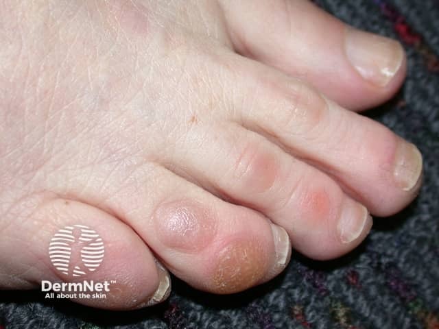
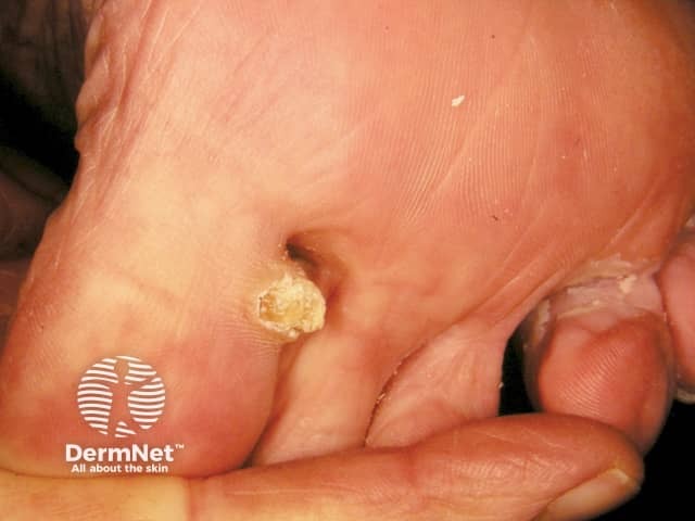
Common keratinocytic lesions
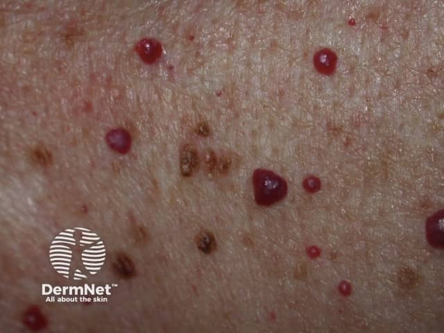
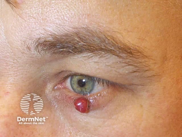
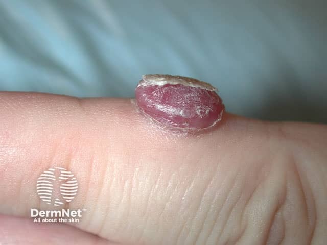
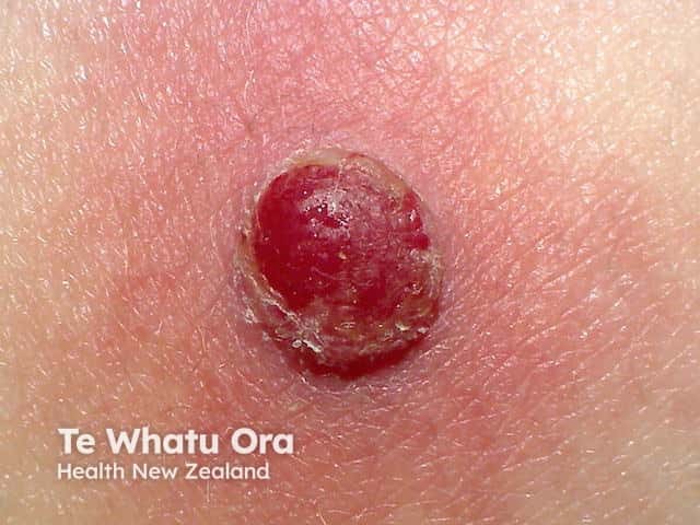
Common lesions of vascular origin
Common fibrous lesions
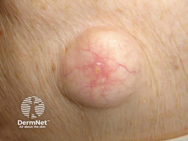
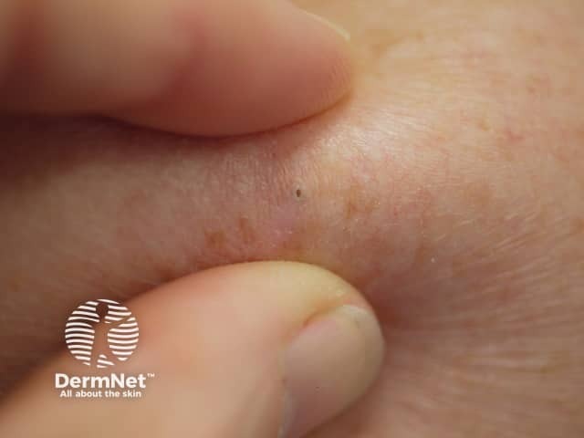
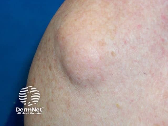
Common subcutaneous lesions
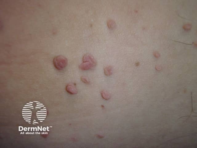
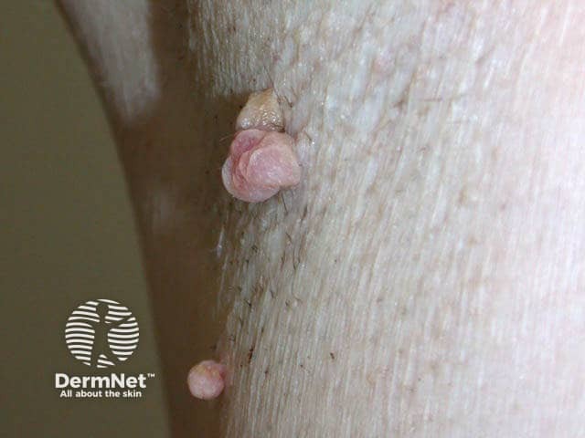
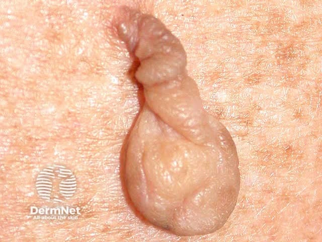
Skin tags
A benign skin lesion is a non-cancerous skin growth.
Any individual from any age group can present with a benign skin lesion.
The features in common for benign skin lesions include:
Benign lesions can be classified by their cellular origin: melanocytic, keratinocytic, vascular, fibrous, fat, and so on,
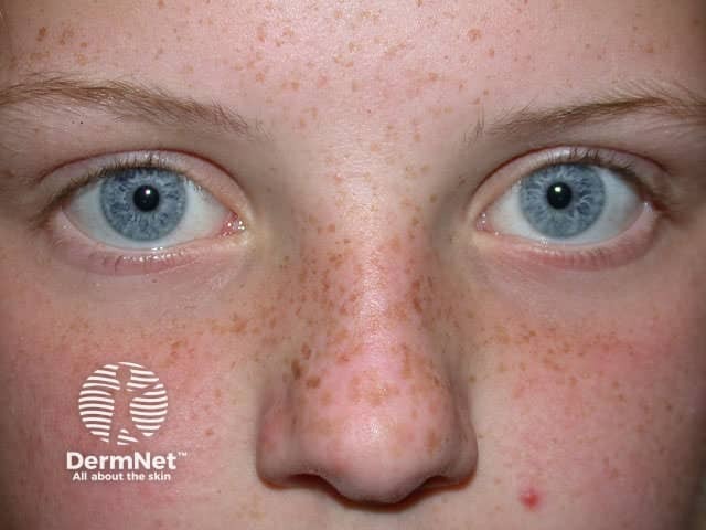
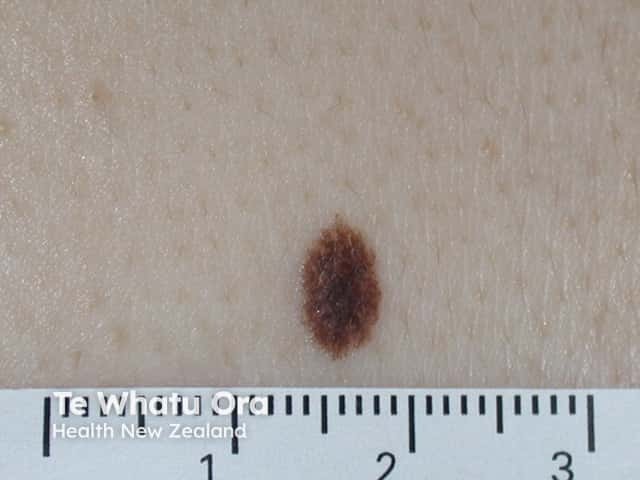
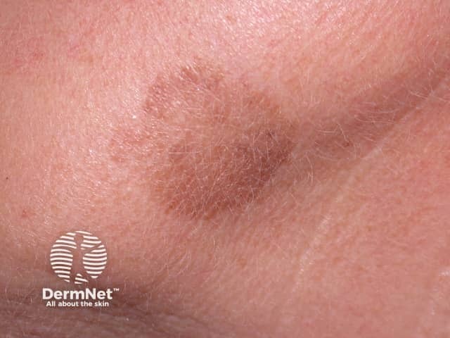
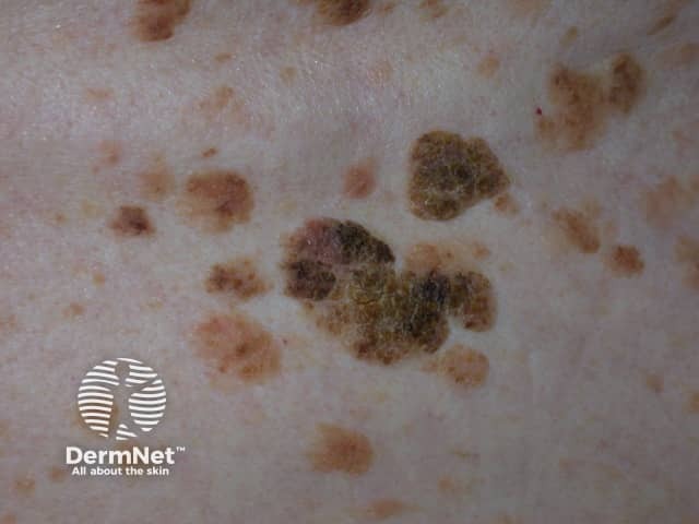
Common benign skin lesions of melanocytic origin include the ephilis, lentigo simplex, and melanocytic naevus (mole).
Ephilides are genetically determined well-defined small brown macules with the following characteristics:
Lentigo simplex is not sun-induced. It has the following characteristics:
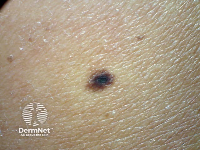
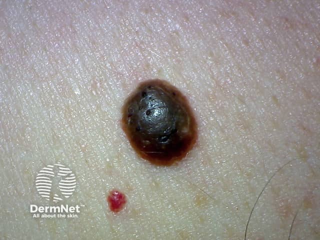
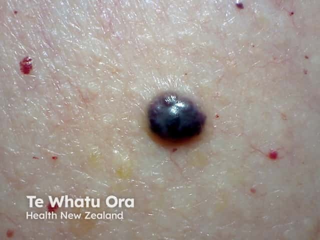
A melanocytic naevus can be histologically classified as a junctional, compound, or dermal naevus depending on the location of nests of naevus cells.
A junctional naevus has naevus cells at the base of the epidermis.
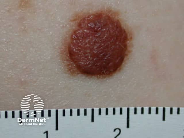
A compound naevus has papular and flat components due to junctional and dermal naevus cells.
A dermal naevus is characterised by naevus cells in the dermis.
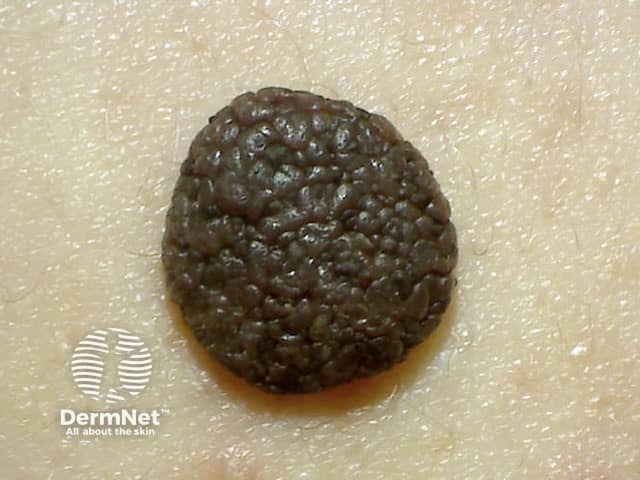
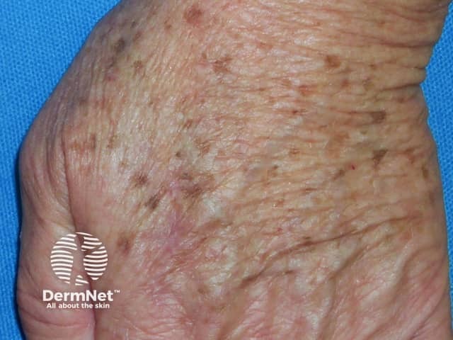
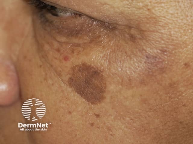
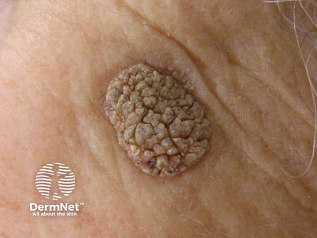
Benign keratoses include solar lentigo and seborrhoeic keratosis.
A solar lentigo is a sun-induced pigmented macule.
Seborrhoeic keratosis presents as a variable warty plaque.
Stucco keratoses are flat-topped keratotic papules.
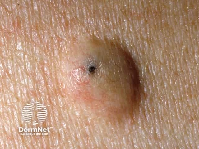
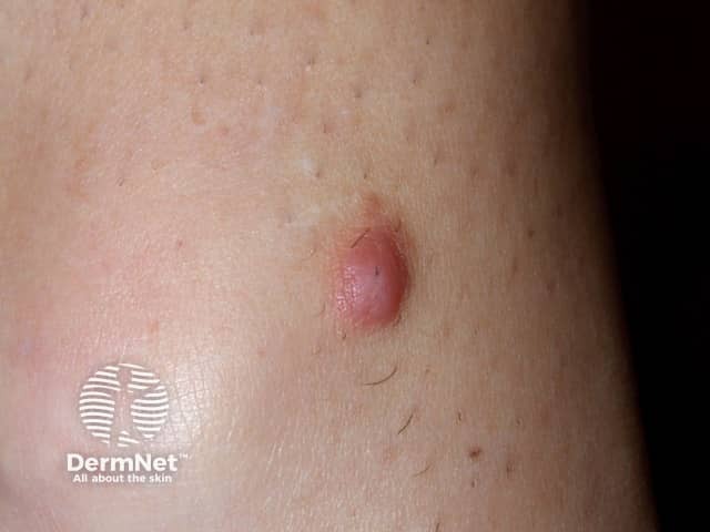
Epidermoid cyst is a follicular nodule with a central punctum.
Corns and calluses are localised areas of thickened skin induced by pressure

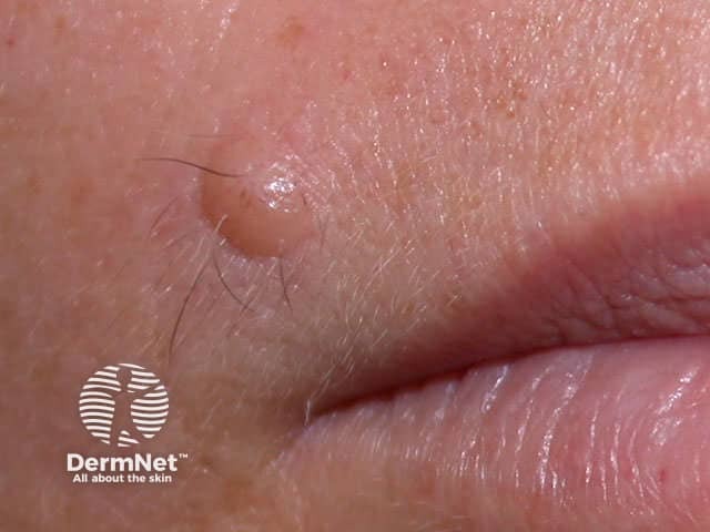
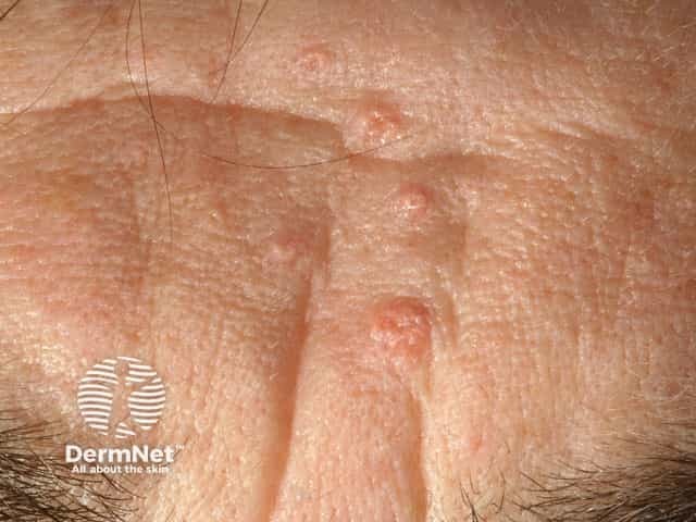
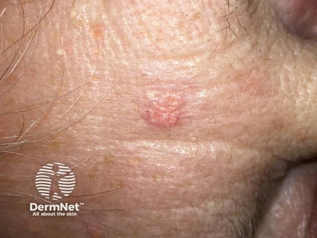
Sebaceous hyperplasia occurs on the forehead and cheeks of adults.

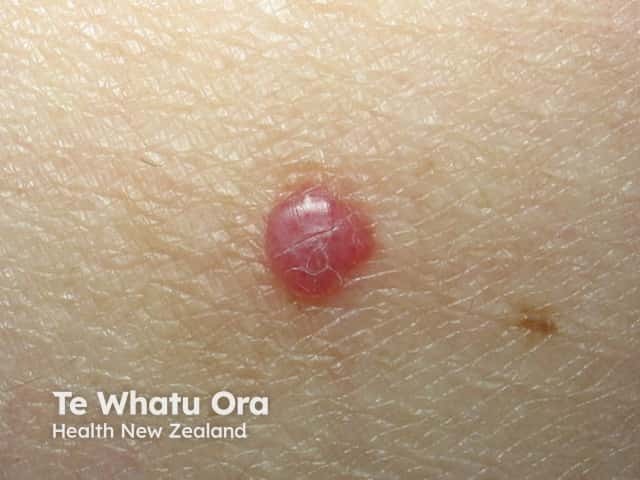
An angioma is due to the proliferation of the endothelial cells.
A pyogenic granuloma is a vascular response to trauma and bacterial infection.
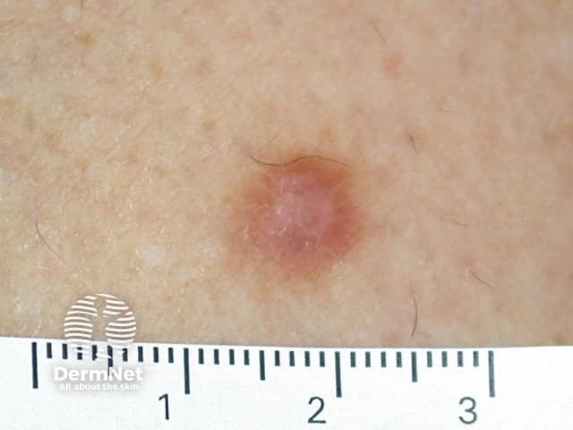
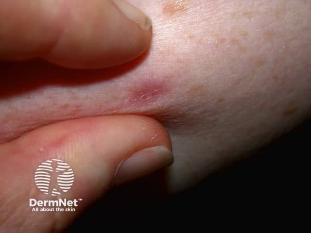
Dermatofibroma is a reactive lesion that presents as one or more firm dermal papules.
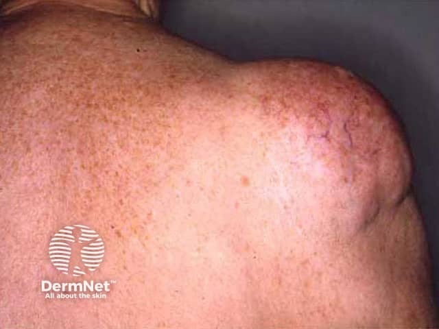
The lipoma is the most common benign soft-tissue tumour.

The most common type of skin tag is also called acrochordon.