

Cutaneous marginal zone lymphoma — extra information
Introduction Demographics Causes Clinical features Diagnosis Differential diagnoses Treatment Outlook
What is primary cutaneous marginal zone lymphoma?
Primary cutaneous marginal zone lymphoma is a type of low-grade cutaneous B-cell lymphoma originating from the mucosa-associated lymphoid tissue (MALT). It is a heterogeneous proliferation of marginal zone cells, B cells, small lymphocytes, and plasma cells.
Who gets primary cutaneous marginal zone lymphoma?
Cutaneous marginal zone lymphoma affects all ages but tends to affect younger patients in comparison to other types of B-cell lymphoma [1]. The median age of diagnosis is 55 years. Men are twice as likely to get this type of lymphoma as women [2].
What causes primary cutaneous marginal zone lymphoma?
It remains unclear whether cutaneous marginal zone lymphoma is the result of a de novo neoplastic process (the beginnings of a tumour) or a reaction to exogenous and/or endogenous agents [3]. A small number of cases have been associated with Borrelia burgdorferi (the cause of Lyme disease), and hepatitis C infection [1–4].
What are the clinical features of primary cutaneous marginal zone lymphoma?
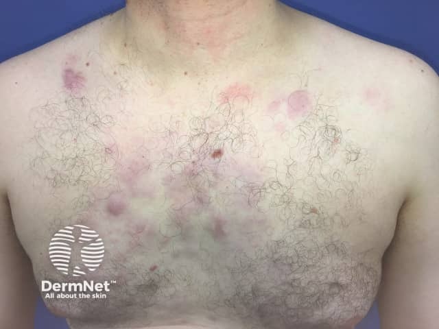
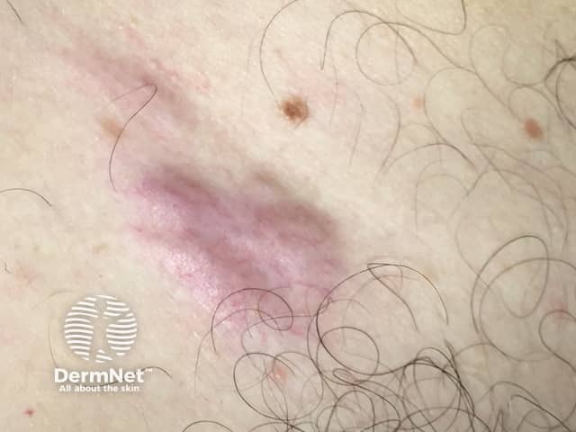
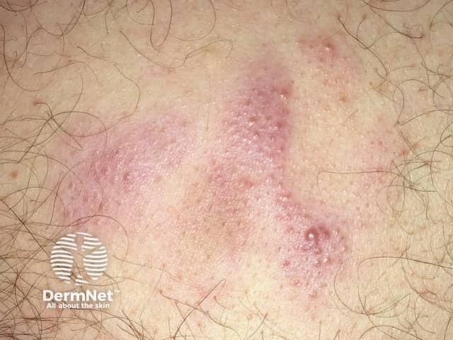
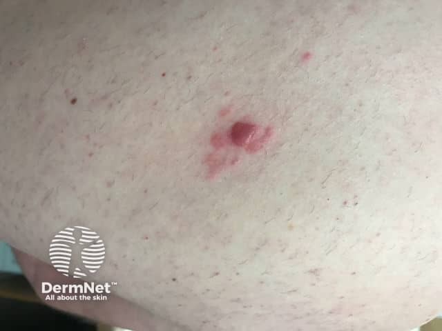
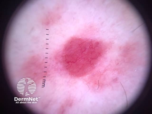
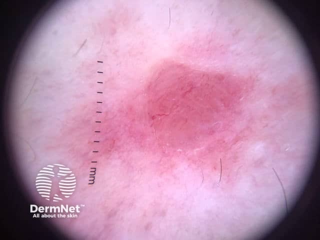
Cutaneous marginal zone lymphoma presents as papules, plaques, or nodules that range in colour from red to violet. The lesions may be solitary or multifocal and are most commonly seen on the patient's trunk or arms [1,2,4]. Dermoscopy is vascular but the features are not specific for the disease.
How is primary cutaneous marginal zone lymphoma diagnosed?
After obtaining a full history from the patient and performing a physical examination and skin examination, the diagnosis is made based on an adequate full-thickness biopsy of the affected skin.
A 4–6 mm punch, incisional, or excisional biopsy should be obtained. The biopsy specimen should include the reticular dermis and fat [2].
Histologically, cutaneous marginal zone lymphomas show nodular to diffuse dermal infiltration of marginal zone B-cells, lymphocytes, lymphoplasmacytoid cells, and plasma cells combined with centroblast- or immunoblast-like cells and reactive T cells. Marginal zone B cells are small to medium in size and have irregular nuclei, inconspicuous nucleoli, and abundant pale cytoplasm [2].
Immunohistochemistry tests of cutaneous marginal zone lymphomas are positive for CD20, CD79a, and BCL-2, and negative for CD 10, and BCL-6 cells [2].
Other essential investigations include a full blood count, comprehensive metabolic profile, and lactate dehydrogenase studies. A test for Borrelia burgdorferi may also be performed, particularly in endemic regions [2].
Additional blood tests that may be performed for the evaluation and staging of the disease include antinuclear antibody, rapid plasma reagin, viral hepatitis serologies, flow cytometry, serum protein electrophoresis, and quantitative immunoglobulin evaluation [2].
Imaging may include positron emission tomography (PET) and/or computed tomography (CT) [2].
Any lymph node with high PET activity, or measuring > 1.5 cm in length by an imaging study, should be biopsied. A bone marrow biopsy is optional [2].
What is the differential diagnosis for primary cutaneous marginal zone lymphoma?
The differential diagnosis for cutaneous marginal zone lymphoma can include:
- Other types of cutaneous B-cell lymphoma
- B cell-rich cutaneous lymphoid hyperplasia
- Cutaneous pseudolymphoma
- Arthropod bites
- Urticaria
- Leukaemia cutis
- Basal cell carcinoma [1,2,4].
It is important to distinguish primary cutaneous marginal zone lymphoma from non-Hodgkin systemic B-cell lymphoma with cutaneous involvement [1,2].
What is the treatment for primary cutaneous marginal zone lymphoma?
Spontaneous resolution of cutaneous marginal zone lymphoma can occasionally occur, particularly in the first few months [2]. A solitary plaque or localised disease can be treated with radiotherapy or surgical excision, with curative intent [1,5].
Multifocal disease can be treated with:
- Observation
- Radiotherapy
- Class 1 topical corticosteroid (eg, clobetasol propionate)
- Nitrogen mustard
- Imiquimod cream
- Cryotherapy
- Intralesional steroids
- Interferon-α
- Rituximab
- Intravenous rituximab
- Chlorambucil [1,5].
Patients who develop disseminated skin lesions are treated with multi-agent chemotherapy [5].
Antibiotic therapy may be indicated in patients who are positive for Borrelia burgdorferi antibodies [5].
What is the outlook for primary cutaneous marginal zone lymphoma?
The prognosis for primary cutaneous marginal zone lymphoma is excellent, with a 5-year survival rate of up to 99% [5]. However, the cutaneous relapse rate is high at approximately 40% [2], but the extracutaneous spread of disease is rare [2].
References
- Sundram U. Primary cutaneous B-cell lymphomas. Surg Pathol Clin. 2014 Jun; 7: 253–83. DOI: 10.1016/j.path.2014.02.002. PubMed
- Suárez AL, Pulitzer M, Horwitz S, Moskowitz A, Querfeld C, Myskowski PL. Primary cutaneous B-cell lymphomas: part I. Clinical features, diagnosis, and classification. J Am Acad Dermatol 2013; 69: 329.e1–13; quiz 341–2. DOI: 10.1016/j.jaad.2013.06.012. PubMed
- Kempf W, Kazakov DV, Mitteldorf C. Cutaneous lymphomas: an update. Part 2: B-cell lymphomas and related conditions. Am J Dermatopathol 2014; 36: 197–208; quiz 209–10. DOI: 10.1097/DAD.0b013e318289b20e. PubMed
- Swerdlow SH. Cutaneous marginal zone lymphomas. Semin Diagn Pathol. 2017 Jan; 34: 76-84. DOI: 10.1097/DAD.0b013e318289b20e. PubMed
- Suárez AL, Querfeld C, Horwitz S, Pulitzer M, Moskowitz A, Myskowski PL. Primary cutaneous B-cell lymphomas: part II. Therapy and future directions. J Am Acad Dermatol 2013; 69: 343.e1–11; quiz 355–6. DOI: 10.1016/j.jaad.2013.06.011. PubMed
On DermNet
- Lymphoma
- Cutaneous B-cell lymphoma
- Skin signs of haematological diseases
- Skin signs and systemic disease
Other websites
- Lymphoma Network New Zealand
- Cutaneous B-cell lymphoma — Lymphoma Research Foundation
- Cutaneous B-cell lymphoma — Medscape
- Lymphoma — Leukaemia and Blood Cancer New Zealand
- Primary cutaneous zone lymphoma — UpToDate
- Clinical manifestations, pathologic features, and diagnosis of extranodal marginal zone lymphoma of mucosa associated lymphoid tissue (MALT) — UpToDate