

Dermoscopy of pityriasis versicolor — extra information
Introduction Clinical features Dermoscopic features Differential diagnoses Histological explanation
What is pityriasis versicolor?
Pityriasis versicolor is a mild chronic infection of the skin caused by lipophilic yeast of the genus Malassezia.
What are the clinical features of pityriasis versicolor?
Pityriasis versicolor presents as discrete or confluent, hyperpigmented, hypopigmented, or pink macules and patches with a fine surface scale, most commonly on the upper trunk. In skin of colour, the infected skin appears paler than the normal surrounding skin.
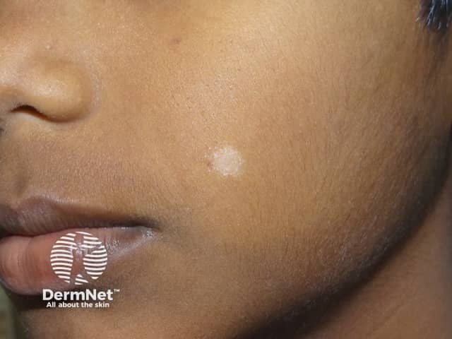
[see also Pityriasis versicolor images]
What are the dermoscopic features of pityriasis versicolor?
Most dermoscopy studies of pityriasis versicolor have been reported in patients with Fitzpatrick IV and V dark skin types. The patches are generally poorly defined, with non-uniform pigmentation and fine white scale. Distribution of the scale can be diffuse or focal, in skin lines, around hair follicles, or at the periphery of the lesion.
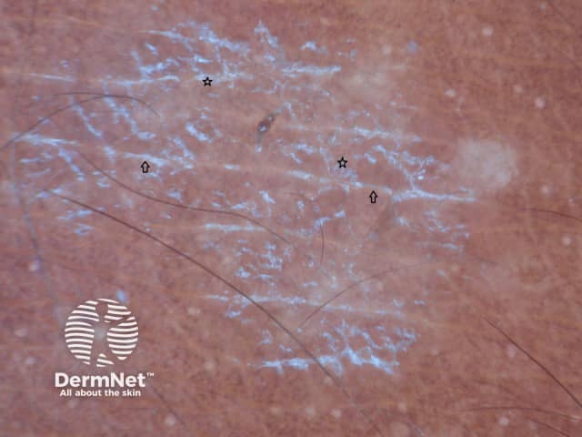
Dermoscopy of hypopigmented pityriasis versicolor shows diffuse white structureless areas with a faint pigment network. A patchy pattern of scale is seen more commonly in hypopigmented pityriasis versicolor compared to the hyperpigmented macules. Fine scales in the natural skin creases can result in a ‘wire-fence’ pattern. On stretching the skin, the scales break into two parts along the skin cleavage lines and are referred to as ‘double edged’ scale.
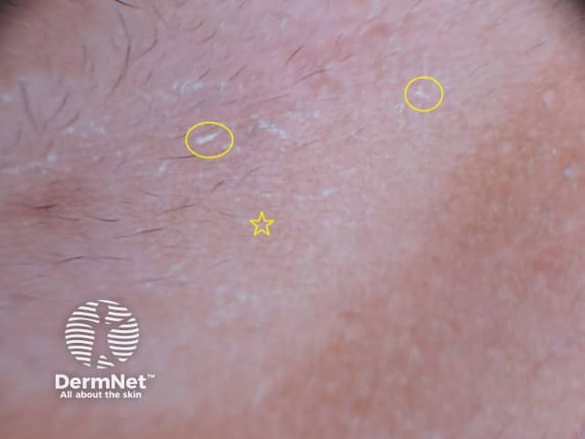
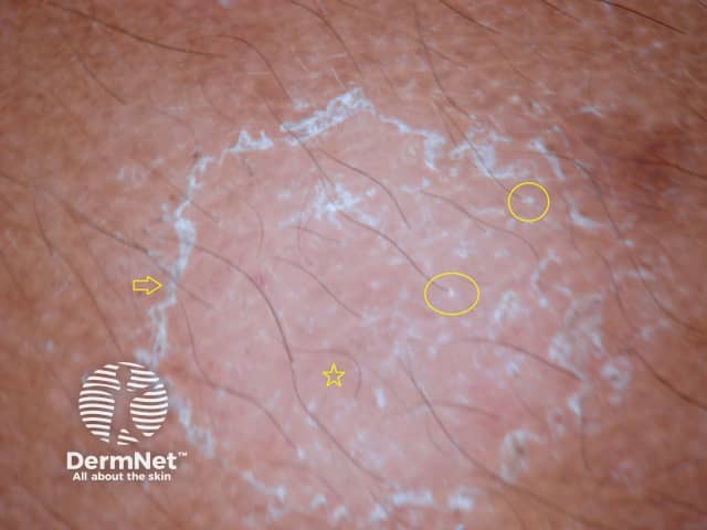
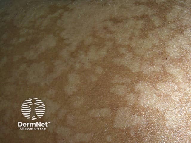
Dermoscopy of hyperpigmented patches of pityriasis versicolor demonstrates diffuse or perifollicular fine whitish scale and a pigmented network of brown stripes or diffuse brownish pigmentation.
What is the dermoscopic differential diagnosis for pityriasis versicolor?
Vitiligo dermoscopy: a milky white structureless area with a diffuse ‘white glow’ due to the absence of a pigment network. Vitiligo has a well-defined border which can be hyperpigmented. The pigment network is usually absent. Hairs within the area are often white with loss of perifollicular pigmentation. [see Dermoscopy of vitiligo]
Pityriasis alba dermoscopy: poorly-defined pale area with fine surface scale scattered in no particular pattern.
Progressive macular hypomelanosis dermoscopy: poorly-defined white macules with faint reticular pigmentation. It is not usually scaly, but white scale is occasionally found with a focal distribution.
What is the histological explanation of the dermoscopic features of pityriasis versicolor?
Skin biopsy of pityriasis versicolor shows mild hyperkeratosis which corresponds with the white scale. Elongated rete ridges have a mild increase in basal layer pigmentation producing the faint background pigment network on dermoscopy [see Pityriasis versicolor pathology].
Bibliography
- Ankad BS. Hypopigmented disorders (disorders of pigmentation). In: Lallas A, Errichetti E, Ionnides D (eds). Dermoscopy in General Dermatology. CRC Press, 2018:39–52.
- Ankad BS, Koti VR. Dermoscopic approach to hypopigmentary or depigmentary lesions in skin of color. Clin Dermatol Rev. 2020:4:79–83. Doi:10.4103/CDR_68_20. Journal
- Bonifaz A, Gómez-Daza F, Paredes V, Ponce RM. Tinea versicolor, tinea nigra, white piedra, and black piedra. Clin Dermatol. 2010;28(2):140–5. doi:10.1016/j.clindermatol.2009.12.004. PubMed
- Errichetti E, Stinco G. Dermoscopy in general dermatology: a practical overview. Dermatol Ther (Heidelb). 2016;6(4):471–507. doi:10.1007/s13555-016-0141-6. PubMed
- Mathur M, Acharya P, Karki A, KC N, Shah J. Dermoscopic pattern of pityriasis versicolor. Clin Cosmet Investig Dermatol. 2019;12:303–9. doi:10.2147/CCID.S195166. PubMed Central
- Thomas N, Malakar S. Dermoscopy: an easy way to solve the diagnostic puzzle in pityriasis versicolor. Indian J Dermatol Venereol Leprol. 2019;85(6):664–5. doi:10.4103/ijdvl.IJDVL_816_16. PubMed
On DermNet
Other websites
Books about skin diseases