Main menu
Common skin conditions

NEWS
Join DermNet PRO
Read more
Quick links
Pigmentary disorders Diagnosis and testing
Author: Prof. Balachandra Ankad, Dermatologist, S. Nijalingappa Medical College, Karnataka, India. DermNet New Zealand Editor in Chief: Adjunct A/Prof Amanda Oakley, Dermatologist, Hamilton, New Zealand. December 2019.
Introduction Dermoscopic features Differential diagnoses Histological explanation
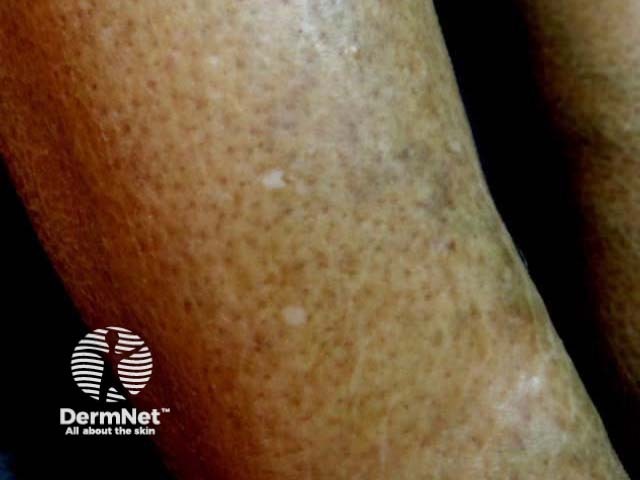
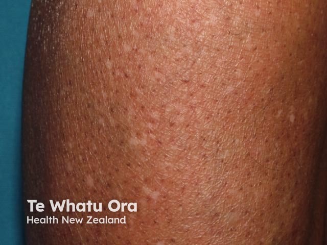
Idiopathic guttate hypomelanosis is a common acquired form of leukoderma characterised by flat porcelain-white macules on sun-exposed areas. The classical sites are the forearms, legs and trunk.
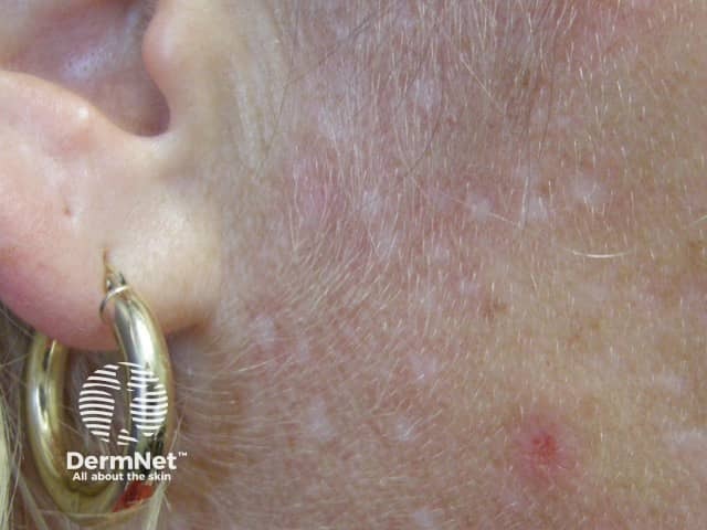
Guttate hypomelanosis
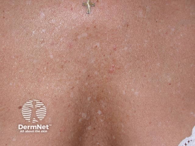
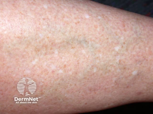
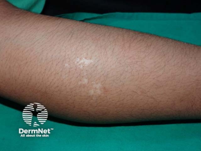
Porcelain-white macules on the lower leg
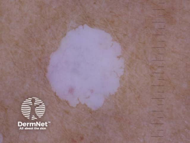
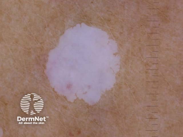
Porcelain-white patch extending peripherally
The dermoscopic features of idiopathic guttate hypomelanosis are characterised by white structureless areas in which the pigment network is absent. They extend peripherally with irregular borders and shapes.
White structureless areas appear to ‘glow’ due to total loss of melanocytes in the epidermis. This glow is not as uniform as the glow seen in the dermoscopy of vitiligo and there may be various shades of white.
Metaphoric terms such as amoeboid, feathery, petaloid and nebuloid shapes have been described [3,4].
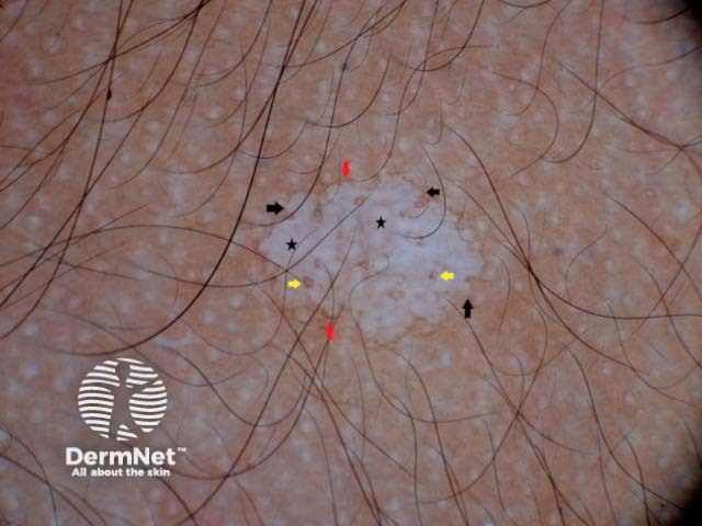
Fig. 1. Amoeboid pattern
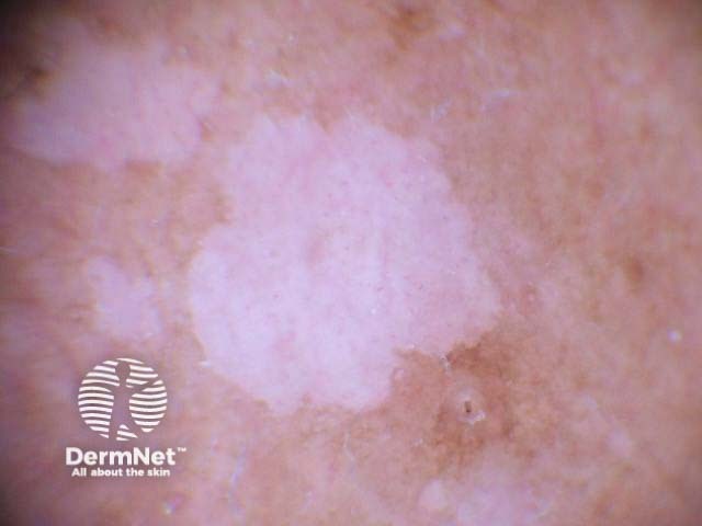
Fig. 2. Amoeboid pattern
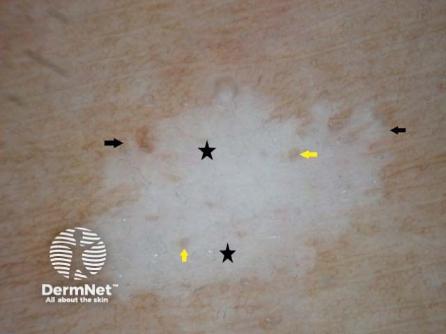
Fig. 3. Feathery pattern
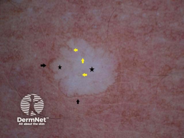
Fig. 4. Petaloid pattern
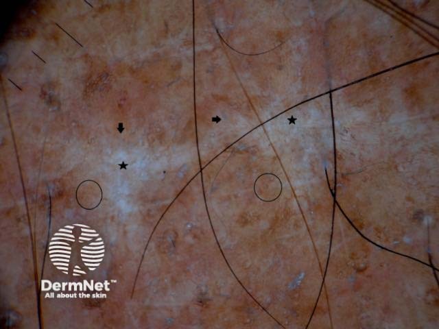
Fig. 5. Nebuloid pattern
Figure legends Figures 1,2. Amoeboid pattern. Diffuse white structureless area (black stars) with well-defined borders which are extending like pseudopods (black arrows), hence the name. Note the perifollicular (yellow arrows) and perilesional (red arrows) pigmentation.
Figure 3. Feathery pattern. White structureless area (black stars) which extends peripherally in linear strands (black arrows) like a feather. Perifollicular pigmentation (yellow arrows) is well appreciated.
Figure 4. Petaloid pattern with a white structureless area (black stars) and peripheral extensions resembling petals. Perifollicular (yellow arrows) and perilesional (black arrows) pigmentation are also noted.
Figure 5. Nebuloid pattern. White areas (black stars) with indistinct borders (black arrows) and subtle pigmentation (black circles).
Macroscopic view Nonpolarised dermoscopy view Polarised dermoscopy view 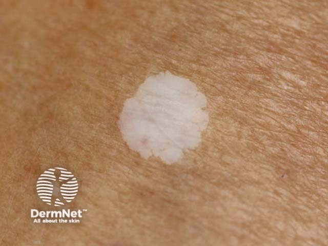
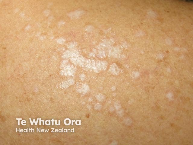
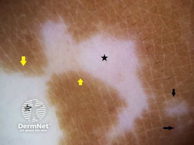
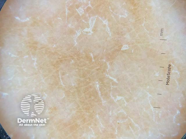
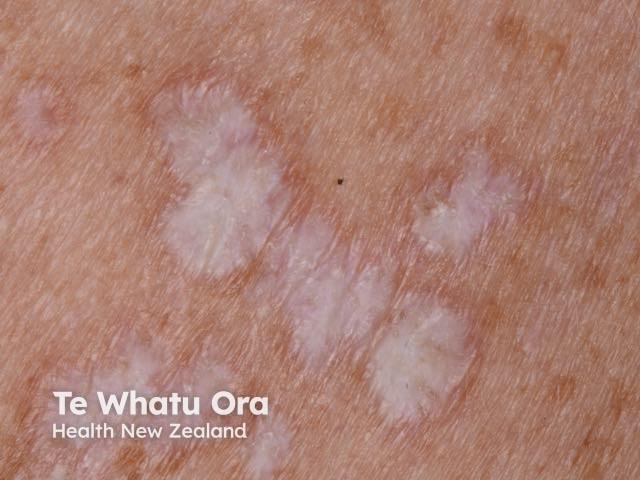
The dermoscopic differential diagnosis for idiopathic guttate hypomelanosis include guttate vitiligo, pityriasis versicolor and lichen sclerosus.
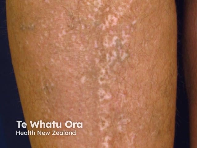
Guttate vitiligo
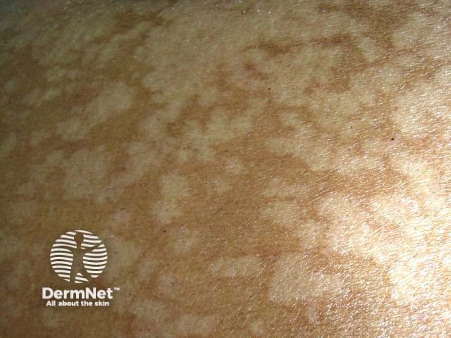
Lichen sclerosus
Dermoscopy of pityriasis versicolor
The histopathology of guttate hypomelanosis shows hyperkeratosis, flattening of rete ridges, epidermal atrophy and acanthosis. There are areas of absent and retained melanocytes; these may explain the white structureless areas and the perifollicular and perilesional pigmentation [5].