

Metastatic melanoma with unknown primary — extra information
Introduction Demographics Causes Clinical features Diagnosis Treatment Outcome
What is metastatic melanoma with unknown primary?
Metastatic melanoma with unknown primary (MUP) refers to metastatic melanoma in lymph nodes, subcutaneous tissue, or visceral sites in the absence of a detectable primary tumour despite detailed examination [1]. Metastatic melanoma should be considered in the differential diagnosis for any patient presenting with a malignancy of unknown origin.
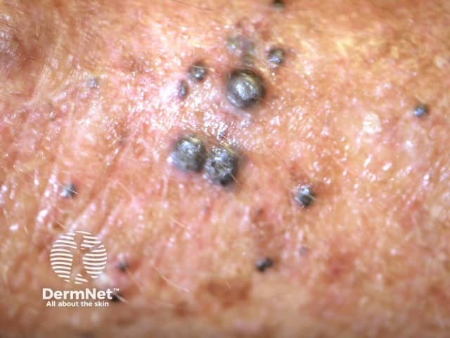
Melanoma commonly metastasises to the regional lymph nodes, liver, lungs, bone, or brain, spreading through lymphatic and haematogenous routes . It can also metastasise to the skin, either locally or to distant sites [2].
For classification purposes, MUP can be separated into subcutaneous, nodal, and visceral disease, with nodal MUP being the most common.


See more images of metastatic melanoma.
Who gets metastatic melanoma with unknown primary?
MUP accounts for approximately 2–9% of cases of metastatic melanoma [3]. The mean age of patients presenting with MUP is in the fifth and sixth decades of life [4]. It more commonly presents in men than in women. This sex difference is currently unexplained.
What causes metastatic melanoma with unknown primary?
The origin of MUP is not fully understood. Some possible explanations are included below.
- Tumour cells at the primary site regressed due to the activity of tumour-infiltrating lymphocytes. Regression in melanoma is well documented, with a frequency of up to 10%.
- The primary cutaneous melanoma may have been excised or otherwise destroyed without adequate pathological analysis.
- The primary melanoma was incorrectly interpreted as being a benign naevus based on clinical and/or pathological features.
- Malignant transformation of ectopic melanocytes in lymph nodes or other organs may have occurred de novo [1,5].
Genotyping of MUP shows a mutational pattern similar to cutaneous melanoma and not the pattern seen in primary melanoma arising in other sites, such as the mucosa or central nervous system. MUP mutations include mutations in the BRAF (particularly the V600E subtype) and NRAS genes [6]. This lends support to the theory that MUP represents metastases from an original primary cutaneous melanoma that may or may not have regressed.
What are the clinical features of melanoma with unknown primary?
As the primary site of the melanoma is unknown, the presentation of MUP is atypical, in that there is no clinically apparent primary cutaneous lesion.
Rarely, the primary melanoma is later found at an extracutaneous site, such as in the eye, or in a sinonasal, vulvovaginal, or gastrointestinal area. In most cases, the primary melanoma cannot be found [6].
The most common clinical presentation of MUP is lymph node disease without clinical or radiological evidence of visceral involvement. Lymph node metastases commonly arise in the axillary (50%), neck (26%), and groin (20%) lymph nodes [1].
In cases of MUP spreading to visceral sites, the initial symptoms are site-specific.
- Hepatic melanoma may present with hepatomegaly, jaundice, or an abdominal mass.
- Pulmonary melanoma can include a pulmonary lesion or pleural effusion.
Advanced MUP that has spread to distant sites may also present with systemic features related to cytokine production, such as fever, weight loss, and anaemia [7].
How is metastatic melanoma with unknown primary diagnosed?
The diagnosis of MUP is usually made based on clinical signs and symptoms consistent with metastatic disease, along with histopathology of a tissue specimen that confirms the presence of malignant melanocytes, such as excisional biopsy of the lymph node or needle core biopsy of a solid organ metastasis [8].
The histological features of MUP on a tissue specimen include:
- The presence of the brown pigment melanin — this is not present in amelanotic melanoma
- Positive melanocytic immunohistochemical markers, such as S100, Melan-A, HMB-45 and SOX10 (SOX10 has been shown to be reliable marker for identifying metastatic melanoma) [8,9].
It is currently not possible to predict the primary site of MUP from histology, immunohistochemistry, or genetics [8].
The requirement of an extensive physical examination to search for the primary lesion has been questioned. Examinations such as ophthalmoscopy (eye examination by ophthalmoscope), otoscopy (ear examination with otoscope), rhinopharyngoscopy (nasal endoscopy and examination of upper airways), laryngoscopy (examination of the inside of the larynx), sigmoidoscopy (rectal examination) and, in women, gynaecological examination, have traditionally been undertaken, yet the yield in finding a primary lesion is not high. These examinations may be costly, time-consuming, and uncomfortable for patients. Special physical examinations are undertaken using clinical judgement in individual cases [4].
Other recommended investigations for MUP include CT imaging of the head, neck, brain (preferably by MRI), chest, abdomen, and pelvis to detect any visceral involvement. CT-PET scanning may also aid in staging the disease.
What is the treatment for metastatic melanoma with unknown primary?
Staging
Studies have shown that lymph node and subcutaneous MUP have a better prognosis than stage III melanoma of a known primary site. The American Joint Committee on Cancer (AJCC) recommends:
- MUP presenting in regional lymph nodes should be classified as stage III rather than stage IV disease
- MUP presenting in visceral sites should be classified as stage IV [1,3].
Treatment
For nodal MUP, radical lymph node dissection of the affected region is generally undertaken. Patients who undergo surgery are less likely to have a recurrence of the malignancy and have improved survival compared to patients undertaking other treatments. Some patients with stage III MUP may benefit from adjuvant systemic and radiation therapy with identical criteria to patients with a known primary melanoma when undergoing the same treatment [1,10].
Subcutaneous MUP behaves more like thick primary melanoma and is generally treated by wide local excision and sentinel lymph node biopsy, if indicated. An epidermal component is sometimes identified in the wide local excision specimen, establishing it as primary cutaneous melanoma rather than MUP [10].
A full metastatic work-up is undertaken for MUP that develops in a visceral site, including cross-sectional imaging. Management involves the resection of any isolated lesion where possible.
While adjuvant therapy has an established role in stage III MUP, its role is unclear in stage IV MUP [10].
What is the outcome for metastatic melanoma of unknown primary?
Results from studies comparing patients with patients with MUP with matched groups of patients with cutaneous melanoma have shown that MUP has similar or better overall survival rates than cutaneous melanoma.
A 2015 systematic review and meta-analysis by Bae et al showed that compared with melanoma of known primary, MUP has a better overall survival, with a hazard ratio of 0.83 for stage III disease and a hazard ratio of 0.85 for stage IV disease [11].
Favourable prognostic factors include:
- Low number of involved lymph nodes
- Female gender
- Absence of visceral metastases (stage IV disease)
- Low serum lactate dehydrogenase (LDH) in those with stage IV disease
- Early surgical intervention [8,12].
Patients with MUP may have improved survival because there may be a more active tumour-directed immune response against the malignant cells (supporting the idea that MUP may be a result of tumour regression) than in melanoma of known primary [13].
References
- Van Beek EJAH, Balm AJM, Nieweg OE, et al. Treatment of regional metastatic melanoma of unknown primary origin. Cancers (Basel). 2015; 7: 1543–53. DOI: 10.3390/cancers7030849. PubMed
- Situm M, Buljan M, Kolić M, Vučić M. Melanoma — clinical, dermatoscopical, and histopathological morphological characteristics. Acta Dermatovenerol Croat. 2014; 22(1): 1–12. PubMed
- Pfeil AF, Leiter U, Buettner PG, et al. Melanoma of unknown primary is correctly classified by the AJCC melanoma classification from 2009. Melanoma Res 2011; 21: 228–34. DOI: 10.1097/CMR.0b013e32834577ec. PubMed
- Tos T, Klyver H, Drzewiecki KT. Extensive screening for primary tumor is redundant in melanoma of unknown primary. J Surg Oncol. 2011: 104; 724–7. DOI: 10.1002/jso.21994. PubMed
- Kibbi N, Kluger H, Choi JN. Melanoma: clinical presentations. In: Kaufman HL, Mehnert JM (eds). Melanoma. Switzerland: Springer; 2016: 107–129
- Egberts F, Bergner I, Kruger S, et al. Metastatic melanoma of unknown primary resembles the genotype of cutaneous melanomas. Ann Oncol 2014; 25: 246–50. DOI: 10.1093/annonc/mdt411. PubMed
- Kamposioras K, Pentheroudakis G, Pectasides D, Pavlidis N. Malignant melanoma of unknown primary site. To make the long story short. A systematic review of the literature. Crit Rev Oncol Hematol. 2011; 78: 112–26. DOI: 10.1016/j.critrevonc.2010.04.007. Journal
- Oien KA. Pathologic evaluation of unknown primary cancer. Semin Oncol. 2009; 36: 8–37. DOI: 10.1053/j.seminoncol.2008.10.009. Journal
- Willis BC, Johnson G, Wang J, Cohen C. SOX10: a useful marker for identifying metastatic melanoma in sentinel lymph nodes. Appl Immunohistochem Mol Morphol. 2015; 23: 109–12. DOI: 10.1097/PAI.0000000000000097. Journal
- Sondak VK, Messina JL. Unusual presentations of melanoma: melanoma of unknown primary site, melanoma arising in childhood, and melanoma arising in the eye and on mucosal surfaces. Surg Clin North Am. 2014; 94: 1059–73. DOI: 10.1016/j.suc.2014.07.010. PubMed
- Bae JM, Choi YY, Kim DS, et al. Metastatic melanomas of unknown primary show better prognosis than those of known primary: a systematic review and meta-analysis of observational studies. J Am Acad Dermatol 2015; 72: 59–70. DOI: 10.1016/j.jaad.2014.09.029. Journal
- Neuman HB, Patel A, Ishill N, et al. A single-institution validation of the AJCC staging system for stage IV melanoma. Ann Surg Oncol 2008; 15: 2034–41. DOI: 10.1245/s10434-008-9915-0. Journal
- Van der Ploeg AP, Haydu LE, et al. Melanoma patients with an unknown primary tumor site have a better outcome than those with a known primary following therapeutic lymph node dissection for macroscopic (clinically palpable) nodal disease. Ann Surg Oncol 2014; 21: 3108–16. DOI: 10.1245/s10434-014-3679-5. Journal
On DermNet
- Metastatic melanoma
- Melanoma pathology
- Cutaneous metastasis
- Melanoma
- Superficial spreading melanoma
- Acral lentiginous melanoma
- Melanoma of nail unit
- Nodular melanoma
- Genetics of melanoma
- Skin cancer
Other websites
- Melanoma New Zealand
- Cancer of unknown primary staging — Medscape
- Melanoma — American Academy of Dermatology