

Angiofibroma — extra information
Introduction Demographics Causes Clinical features Complications Diagnosis Differential diagnoses Treatment Outcome
What is an angiofibroma?
A cutaneous angiofibroma is a benign vascular neoplasm composed of dermal fibrous tissue and blood vessels.
Angiofibroma is classified by association with a genetic disorder or according to its body site [1].
Who gets angiofibromas?
Angiofibromas are associated with the following genetic disorders:
- Tuberous sclerosis
- Birt-Hogg-Dubé syndrome
- Multiple endocrine neoplasia type 1 (MEN-1).
Angiofibromas are more commonly acquired.
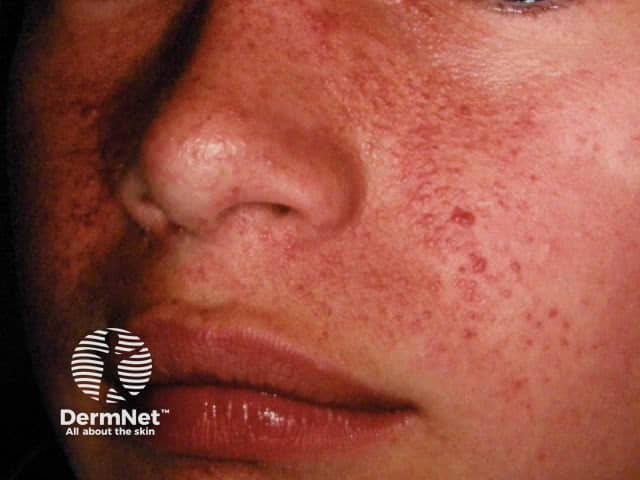
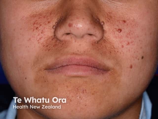
Tuberous sclerosis
Tuberous sclerosis is a neurocutaneous autosomal dominant syndrome, in which angiofibromas appear in childhood in the nasolabial folds and on the central face [2]. Patients with tuberous sclerosis commonly develop an oral fibroma or a periungal angiofibroma (Koenen tumour) over time [1]. The facial angiofibromas associated with tuberous sclerosis are also called adenoma sebaceum, juvenile angiofibroma, and Pringle tumour.

Birt-Hogg-Dubé syndrome
Facial angiofibromas have been reported in Birt-Hogg-Dubé syndrome, a rare genodermatosis characterised by skin and renal tumours, as well as spontaneous pneumothorax [3]. Most of the cutaneous lesions however are fibrofolliculomas, which are abnormal growths of the hair follicles.
Multiple endocrine neoplasia type 1
Multiple endocrine neoplasia type 1 is a hereditary syndrome that leads to tumours in several endocrine organs [1].
Acquired angiofibroma
Angiofibromas can also be acquired and unrelated to a genetic syndrome, commonly in the form of:
- Fibrous papule of the nose/face
- Pearly penile papules.
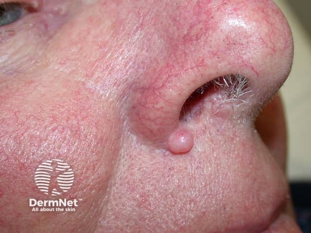
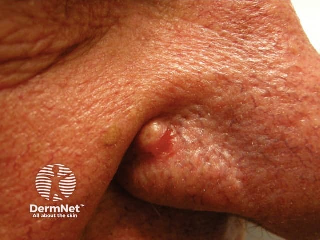
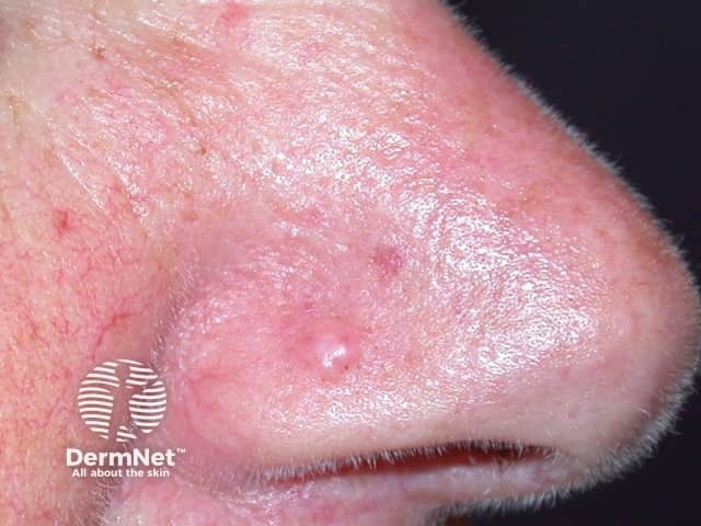
A fibrous papule is characteristically found in adults as a solitary lesion usually on the nose, often clinically mistaken for a basal cell carcinoma or melanocytic naevus. It is thought to be a form of dermal naevus.
Multiple pearly penile papules occur in 10–30% of adult males on the coronal edge and sulcus. They can be mistaken for viral warts [1,2].
See penile pearly papules images.
What causes angiofibromas?
Angiofibromas are caused by a local overgrowth of collagen, fibroblasts, and blood vessels.
- In tuberous sclerosis, mutations are present in tuberous sclerosis complex 1 (TSC1), which encodes the protein hamartin, and tuberous sclerosis complex 2 (TSC2) which encodes the protein tuberin.
- Birt-Hogg-Dubé syndrome is due to a mutated FLCN gene, which encodes the protein folliculin [3].
- Multiple endocrine neoplasia type 1 results from a mutation in the MEN1 gene which produces the protein menin [1].
Genetic mosaicism for these genetic conditions must also be considered [4]. What specifically triggers the development of angiofibroma is unknown.
What are the clinical features of angiofibroma?
An angiofibroma is a firm, flesh-coloured dome-shaped papule less than 5 mm in diameter. Small capillaries may be visible on the surface of the lesion.
- Facial angiomas associated with a genetic syndrome are commonly found in clusters in the butterfly region of the face.
- A fibrous papule of the face is usually a solitary lesion located on the nose in an adult.
- Pearly penile papules are 1–3 mm skin coloured or white papules in a row along the penile coronal margin [5].
What are the complications of angiofibromas?
Angiofibromas may be itchy and may also bleed. Those associated with genetic syndromes result in facial disfigurement and stigmatisation [1]. See Psychosocial factors in dermatology.
How are angiofibromas diagnosed?
The diagnosis of angiofibroma may be made clinically or after a skin biopsy. The histopathology of angiofibroma shows an ‘onion skin’ pattern around vessels and follicles, hyperkeratosis, and vascular proliferation [5].
If an underlying genetic condition is suspected, appropriate genetic screening and evaluation are required [1].
What is the differential diagnosis for angiofibromas?
The differential diagnosis for angiofibroma depends on its location [1].
Differential diagnoses for facial lesions that can resemble angiofibromas can include:
- Intradermal melanocytic naevus
- Acne
- Basal cell carcinoma
Differential diagnoses for periungual lesions that can resemble angiofibroma can include:
Differential diagnoses for penile lesions that can resemble angiofibroma can include:
What is the treatment for angiofibroma?
Angiofibromas are benign and do not always require removal. Options for treatment of angiofibromas include:
- Excision
- Dermabrasion
- Using lasers, electrical, and radiofrequency devices
- Cryotherapy
- Topical podophyllotoxin
- Topical rapamycin
- Topical beta-blocker (eg, timolol) [6].
Multiple treatments are often necessary [1].
What is the outcome for angiofibromas?
Although angiofibromas are benign, they are persistent. Angiofibromas can be removed for cosmetic or pain-related reasons. The recurrence rate for angiofibromas associated with tuberous sclerosis may be as high as 80% [1].
References
- Macri A, Tanner LS. Cutaneous Angiofibroma. StatPearls; Treasure Island: StatPearls Publishing; 2020. PubMed
- Billings SD, Goldblum JR. Soft tissue tumors and tumor-like reactions. In: Dermatopathology. Elsevier Inc.; 2010: 499–564. doi: 10.1016/B978-0-443-06654-2.00013-5.
- Schaffer JV, Gohara MA, McNiff JM, Aasi SZ, Dvoretzky I. Multiple facial angiofibromas: a cutaneous manifestation of Birt-Hogg-Dubé syndrome. J Am Acad Dermatol. 2005;53(2 Suppl 1):S108-11. doi:10.1016/j.jaad.2004.11.021. PubMed
- Cohen BA. Nodules and tumors. In: Pediatric Dermatology: Fourth Edition. Elsevier Inc.; 2013: 126–47. doi: 10.1016/B978-0-7234-3655-3.00005-9.
- Johnston RB. Fibrous tumors and tumor-like proliferations. In: Weedon’s Skin Pathology Essentials. Elsevier; 2017: 612–39. doi:10.1016/b978-0-7020-6830-0.50034-7.
- Krakowski AC, Nguyen TA. Inhibition of angiofibromas in a tuberous sclerosis patient using topical timolol 0.5% Gel. Pediatrics. 2015;136(3):e709-13. doi:10.1542/peds.2015-0025. Journal
On DermNet
- Angiofibroma pathology
- Angiofibromas images
- Tuberous sclerosis
- Tuberous sclerosis images
- Skin signs of neurological diseases
- Skin lesions, tumours and cancers
- Vascular skin problems
- Facial skin problems
Other websites
- Angiofibroma clinical images at Medicine Net
- Online Mendelian Inheritance in Man