

Onychogryphosis — extra information
Introduction Demographics Causes Clinical features Complications Diagnosis Differential diagnoses Treatment Outcome
What is onychogryphosis?
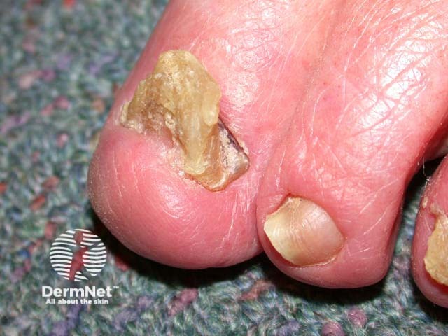
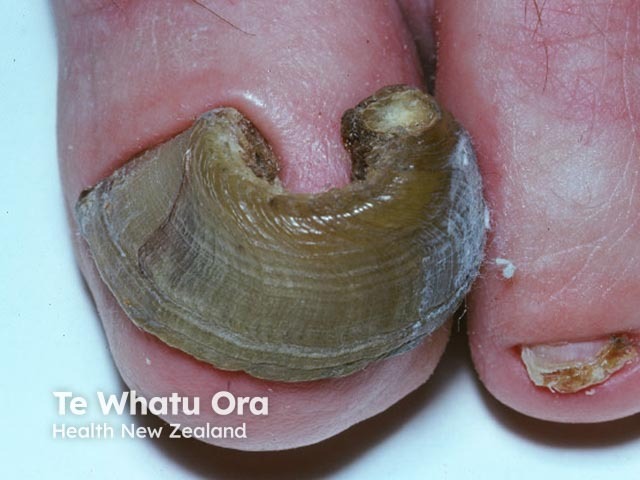
Onychogryphosis, also known as ram’s horn nail, is a nail disorder resulting from slow nail plate growth. It is characterised by an opaque, yellow-brown thickening of the nail plate with elongation and increased curvature [1,2].
Who gets onychogryphosis?
There have been a few reports of congenital onychogryphosis, and onychogryphosis can be seen in a number of rare genodermatoses.
Acquired onychogryphosis is more common [1]. It is more often observed in older people, people with poor personal care, and patients with senile dementia [1–3].
What causes onychogryphosis?
The exact cause of onychogryphosis is not completely known.
It is associated with:
- Skin diseases such as ichthyosis, psoriasis, pemphigus, tertiary syphilis, hyperuricaemia (the cause of gout), and, in the past, smallpox
- Poor peripheral circulation, which may be associated with diabetes mellitus
- Traumatic injury to the nail bed
- Microtrauma due to poorly fitting shoes
- Hallux valgus (bunion) [1].
In onychogryphosis, the nail plate becomes hypertrophied and uneven at the proximal matrix (the nail growth plate). The direction of the deformity is determined by which side grows faster, whether due to an insufficiency of the nail matrix under the proximal nail fold or because the nail bed produces a greater quantity of keratin than normal [1].
What are the clinical features of onychogryphosis?
The clinical features of onychogryphosis include:
- Involvement of one or both great toenails, but any of the nails can be involved
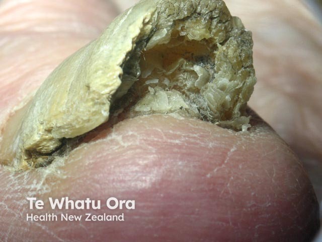
- Opaque, yellow–brown thickening of the nail plate with elongation and increased curvature
- What is often described as a ‘ram’s horn’ or ‘oyster-like’ appearance
- The nail plate initially growing upwards and deviating in a lateral direction towards the other toes
- The nail bed exhibiting an irregular surface marked by striations that are most commonly transverse rather than longitudinal.
What are the complications of onychogryphosis?
The complications of onychogryphosis may include:
- An ingrown toenail
- Paronychia
- Secondary onychomycosis (fungal nail infection)
- Subungual gangrene (rarely).
How is onychogryphosis diagnosed?
Onychogryphosis is diagnosed clinically based on its characteristic appearance [1]. In the early stages, it may be difficult to recognise, as the only feature is hypertrophy of the nail plate, and the classical features usually appear in the later stages [1].
On histology, the keratinocytes appear disorderly and there is associated hyperchromatism, parakeratosis, and numerous splits [1].
What is the differential diagnosis for onychogryphosis?
The differential diagnosis for onychogryphosis includes:
- Congenital malalignment of toenails (where the nail plate is laterally deviated and not parallel to the major axis of the distal phalanx, in infancy or childhood) [4]
- Pachyonychia congenita, in which the fingernails are more commonly affected, and the thickened nail has a brownish discolouration (molecular genetic studies can be done to detect mutations in keratin genes KRT6a and KRT16)
- Onychomycosis, which can co-exist with onychogryphosis (nail clippings should be undertaken to culture dermatophyte fungus, yeast, or mould) [5].
What is the treatment for onychogryphosis?
Treatment for onychogryphosis can be either conservative or operative, depending on its cause and symptoms [1].
Excessive pressure or microtrauma to the nail bed can be minimised by selecting properly fitted footwear [1].
Conservative treatment involves:
- Regular use of an electric drill, bur, or mechanical debridement with a nail clipper to shorten the nail and remove subungual hyperkeratosis
- Cryotherapy prior to debridement will soften the nail plate so it is easier to cut
- Blunt dissection with a nail clipper after medical nail avulsion with either 40% urea or 50% potassium iodide [1].
If conservative treatment fails, nail avulsion may be considered followed by ablative or excisional matricectomy (surgically or chemically removing the proximal nail matrix at the base of the nail) [1].
- Excisional techniques include scalpel excision, cutting electrosurgery, or laser in cutting mode [1].
- Ablative techniques include chemical cautery, electrosurgery, or laser in ablative mode [1].
The Zadik technique or a V–Y advancement flap can be used to completely remove the nail matrix [1]. The Syme method, whereby half of the terminal phalanx is removed with the nail fold, is rarely used [1].
What is the outcome for onychogryphosis?
Onychogryphosis tends to recur after conservative treatment [1]. For both clinical and cosmetic purposes, treatment can be repeated to keep the nail bed short and prevent secondary complications. The use of proper footwear to prevent excessive nail pressure on the nail bed is important.
References
- Ko D, Lipner SR. Onychogryphosis: case report and review of the literature. Skin Appendage Disord 2018; 4: 326–330. doi: 10.1159/000485854. PubMed
- Chang P. Onychogryphosis. Our Dermatology Online 2011; 2: 227–8. Journal
- Chang P, Meaux T. Onychogryphosis: a report of ten cases. Skinmed 2015; 13: 355–9. PubMed
- Cataflo P, Musumec ML, Lacarrubba F, Dinotta F, Micali G. Congenital malalignment of the great toenails: a review. Skin Appendage Disord 2018; 4: 230–5. DOI: 10.1159/000484943. PubMed
- Singh G. Nails in systemic disease. Indian J Dermat Venereol Leprol 2011; 77: 646–51. DOI: 10.4103/0378-6323.86472. Journal
On DermNet
- Ingrown toenail
- Paronychia
- Fungal nail infections
- Nail psoriasis
- Nail disorders
- Medical nail avulsion
- Pachyonychia congenita
- Ichthyosis