

Pachydermodactyly — extra information
Introduction Demographics Causes Clinical features Differential diagnoses Diagnosis Treatment Outcome
What is pachydermodactyly?
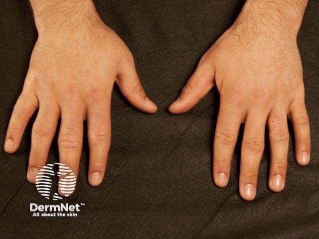
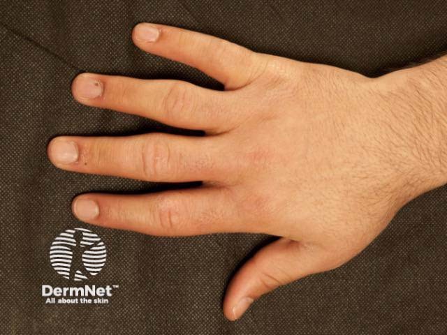
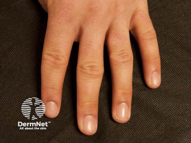
Pachydermodactyly is a benign digital fibromatosis presenting as a progressive asymptomatic periarticular thickening, most commonly around the proximal interphalangeal joints. Palmar and distal interphalangeal (DIP) or metacarpophalangeal (MCP) joint involvement has been described.
Who gets pachydermodactyly?
Pachydermodactyly most commonly occurs in adolescents and young men. The affected male to female ratio is 4:1 [1,2].
What is the cause of pachydermodactyly?
The exact cause of pachydermodactyly is not fully understood. It has been linked to repetitive mechanical stimulation in genetically predisposed individuals. Hormonal factors may also have an influence as illustrated by the predominance in male individuals and symptoms appearing at the age of puberty.
Common triggers identified have been habits such as interlacing, stretching, rubbing and cracking of the fingers, and increased mechanical irritation related to computer use, work, professional sport, and musical activities.
Associations with tuberous sclerosis and Ehlers-Danlos syndrome are reported [1,2].
The pathophysiology is thought to be due to deposition of abnormal collagen in the dermis, especially loose strands of type III and type V collagen.
What are the clinical features of pachydermodactyly?
Pachydermodactyly presents as a symmetrical soft-tissue thickening of the skin on the sides of the proximal interphalangeal joints, most commonly affecting the second (index), third (middle) and fourth (ring) fingers of both hands. The thumbs and fifth fingers are rarely involved. Some patients may also have overlying hyperkeratosis, lichenification, and hyperpigmentation. Movement of the fingers is not restricted. Pachydermodactyly is not associated with symptoms such as pain or stiffness. There is no evidence of a synovitis.
The toes are never affected.
What is the differential diagnosis for pachydermodactyly?
Other conditions that may be considered in the differential of diagnosis of pachydermodactyly include:
- Knuckle pads
- Pseudoknuckle pads.
The lack of symptoms and other signs make the following conditions unlikely:
- Rheumatoid arthritis
- Psoriatic arthritis
- Juvenile idiopathic arthritis
- Microgeodic disease
- Early systemic sclerosis
- Sclerodactyly
- Primary and secondary pachydermoperiostosis.
How is pachydermodactyly diagnosed?
Diagnostic criteria have been proposed [2]:
- no symptoms
- no morning stiffness of the involved joints
- no pain or tenderness related to the involved joints
- swelling involves the sides of the fingers, and does not extend around the fingers
- normal/negative laboratory tests for arthritis
- plain X-rays show only soft tissue swelling.
Investigations for inflammatory arthropathies may include:
- Rheumatoid factor (RF)
- Anti-cyclic citrullinated peptide antibody (Anti-CCP)
- Antinuclear antibody (ANA)
- Extractable nuclear antigens (ENA)
- C-reactive protein (CRP)
- Plain radiograph, to assess soft tissue swelling, periarticular osteopenia, periostosis, erosions, cysts, or osteophytes
- Ultrasound scan, to look for synovial hypertrophy, vascularity, and bony erosions.
Features seen in skin biopsies include [1,3]:
- Increased fibroblast proliferation
- Abnormal collagen deposition in the mid and deep dermis
- Increased mucin deposition around the collagen fibres entrapping the eccrine sweat glands
- Sometimes, hyperkeratosis, and acanthosis.
Inflammation is typically absent.
What is the treatment for pachydermodactyly?
Pachydermodactyly may reverse or resolve with cessation of mechanical stimulation if any.
Intralesional corticosteroid (triamcinolone) and surgical resection have been trialled in severe cases with some benefit.
What is the outcome for pachydermodactyly?
Overall, pachydermodactyly remains stable. Permanent joint deformities or damage are rarely observed.
References
- Dallos T, Oppl B, Kovács L, Zwerina J. Pachydermodactyly: a review. Curr Rheumatol Rep. 2014;16(9):442. doi:10.1007/s11926-014-0442-7. PubMed
- Paravina M, Stanojević M, Jovanović D, Ljubisavljević D. Pachydermodactyly: a case report and literature review. Serbian J Dermatol Venereol. 2014; 6: 174–85. doi: 10.2478/sjdv-2014-0015. Available at: content.sciendo.com/view/journals/sjdv/6/4/article-p174.xml?lang=en (accessed 13 October 2019)
- Barnes LA, Bae GH, Lewis MA, Rieger KE. Pachydermodactyly: Case report including clinical and histopathologic diagnostic pitfalls. J Cutan Pathol. 2018;45(12):949–53. doi:10.1111/cup.13359. PubMed
On DermNet