Introduction
Demographics
Causes
Clinical features
Complications
Diagnosis
Differential diagnoses
Treatment
Outcome
Reactive infectious mucocutaneous eruption (RIME) is a mucocutaneous disease characterised by predominantly mucosal involvement. It commonly follows a bacterial or viral respiratory tract infection and is mostly observed in children and adolescents, although it can occur in adults.
In 2015, the term “mycoplasma pneumoniae-induced rash and mucositis” (MIRM) was introduced to differentiate specific mucocutaneous eruptions from erythema multiforme, Stevens-Johnson syndrome (SJS), and toxic epidermal necrolysis (TEN). The MIRM classification, as originally proposed by Ramien et al. in 2020, has subsequently been modified to “reactive infectious mucocutaneous eruption” (RIME), a broader term that includes non-mycoplasma pneumoniae pathogens capable of causing a clinically similar presentation.
Key characteristics of RIME include:
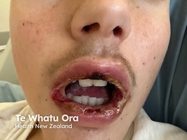
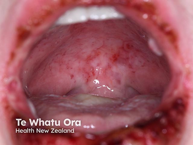
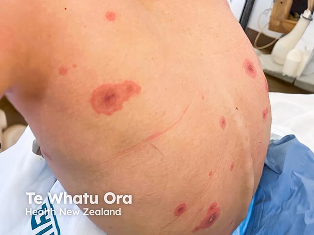
The clinical course of RIME characteristically begins with respiratory symptoms approximately one week prior to the onset of mucocutaneous eruption. Mucositis can affect the mouth, eyes, urogenital organs, and gastrointestinal tract, with lesions tending to be primarily vesiculobullous or targetoid. Cutaneous lesions typically affect the extremities, including acral areas.
RIME typically affects children and adolescents (median age of 12 years) with a slight male predominance; however, rare cases have been reported in adults. A 2019 prospective cohort study showed that 22.7% of children with MP-associated community-acquired pneumonia developed mucocutaneous lesions, with 6.8% developing RIME.
Genetic susceptibility has been suspected following reports of recurrent RIME among affected families. However, specific genetic determinants have not been identified.
There appears to be no association with race or ethnicity.
Although the pathophysiology of RIME is not known, it is hypothesised to involve pathogenic factors and inflammatory response mechanisms. Respiratory infections have been identified to cause RIME, with Mycoplasma pneumoniae being the most frequently associated pathogen.
In addition to M. pneumoniae, other associated pathogens include:
RIME is characterised by severe mucositis and generally sparse, variable cutaneous involvement. Some patients may present with only mucosal involvement, termed RIME “sine rash” (without rash).
Antecedent symptoms, such as fever, malaise, and cough, typically occur one week prior to the onset of mucocutaneous eruption and likely reflect the precipitating infection rather than a true prodromal phase of RIME.
RIME predominantly affects mucous membranes:
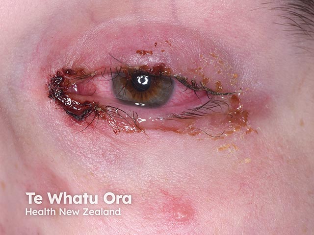
Mucocutaneous complications occur in about 10% of patients, with ocular complications being the most serious. These include:
Cutaneous complications include post-inflammatory pigmentary alteration (eg, hyperpigmentation) and scarring.
Systemic complications are exceedingly rare and include restrictive lung disease, chronic obliterative bronchitis, and B-cell lymphopenia.
Diagnosis is made clinically based on history and physical examination. RIME should be suspected in children or young adults with mucocutaneous eruption that appears about one week following flu-like symptoms, such as fever, malaise, and cough.
Clinical, radiographic, and laboratory evidence of an infectious trigger supports the diagnosis and may include:
Histologic features include apoptotic keratinocytes and sparse perivascular dermal infiltrates. However, biopsy is not routinely performed as histology does not reliably distinguish RIME from other similar mucocutaneous diseases, such as erythema multiforme or SJS/TEN.
There are no established treatment guidelines. However, a comprehensive approach includes supportive measures, early consultation with appropriate specialty departments, and antimicrobial and immunomodulatory therapies as indicated.
Approximately 4% of patients with RIME require admission to intensive care, however this is primarily due to airway complications of M. pneumoniae infection rather than mucocutaneous complications. Ophthalmologic consultation should be sought if any ocular involvement is identified given the risk for serious sequelae.
Antimicrobials:
Immunomodulatory agents:
Systemic corticosteroids and other therapies have been used in the treatment of RIME, often where there is extensive mucosal involvement or high symptom burden. However, high-quality evidence and standard dosing recommendations are currently lacking.
Systemic corticosteroids. Example regimens include:
Steroid-sparing agents. Example regimens include:
Considerations for specific organ involvement:
RIME is associated with a low mortality rate (2-3%) and most patients achieve full recovery. Mucosal synechiae and pigmentary changes are the most common sequelae. Recurrence has been reported in approximately 8% of cases.
Understanding Reactive Infectious Mucocutaneous Eruption (RIME) — The Dermatology Digest

