DermNet provides Google Translate, a free machine translation service. Note that this may not provide an exact translation in all languages

Translate

Chondrodermatitis nodularis

Author: Dr Anita Eshraghi, Dermatologist, Sweden. DermNet NZ Editor-in-Chief Adjunct A/Prof Amanda Oakley, Dermatologist, Hamilton, New Zealand. Original page by Dr Oakley was published in 1997. Updated August 2018.


toc-icon

What is chondrodermatitis nodularis?

Chondrodermatitis nodularis is a common inflammatory condition which affects the skin and cartilage of the helix or antihelix of the ear.

Chondrodermatitis nodularis is sometimes called Winkler disease, after the dermatologist who described it in 1915. It is also called nodular chondrodermatitis, chondrodermatitis nodularis helicis, and chondrodermatitis nodularis chronica helicis.

Who gets chondrodermatitis nodularis?

Chondrodermatitis nodularis occurs more frequently in fair-skinned and middle-aged older males, with 10–35% of the cases reported in women. It is rarely reported in children.

What causes chondrodermatitis nodularis?

The exact cause of chondrodermatitis nodularis is still unknown. There are several factors that contribute to its development.

Chondrodermatitis nodularis

What are the clinical features of chondrodermatitis nodularis? 

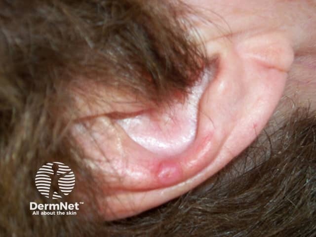
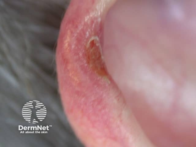
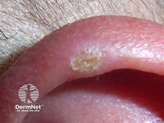
Chondrodermatitis nodularis is a solitary, firm, and oval-shaped nodule, 4–6 mm in diameter, with central crust and surrounding erythema.

  • In men, the most common site for CNH is the helix, while in women it is more often found on the antihelix. 
  • It is typically unilateral, located on the sleeping side, but can be bilateral.
  • CNH is painful and tender. Pain at night may prevent sleeping on the affected side.
  • The lesion can bleed or discharge a small amount of scaly material. 

How is chondrodermatitis nodularis diagnosed?

In most cases, the diagnosis of chondrodermatitis nodularis is made clinically, based on the characteristic location on the helix or antihelix, and a typical history of pain and tenderness.

Sometimes an excision biopsy may be necessary to confirm the diagnosis (see Chondrodermatis nodularis pathology).

What is the differential diagnosis for chondrodermatitis nodularis?

The differential diagnosis for CNH depends on the clinical findings. Other diagnoses to consider include:

What is the treatment for chondrodermatitis nodularis?

General measures

Protective padding at night can relieve pressure on the area affected by CNH.

  • Try to avoid sleeping on one side only.
  • Select a soft pillow.
  • Consider fashioning a ‘hole’ in the pillow to avoid pressure on the painful area.
  • Foam rubber or a bath sponge can be used to make a chondrodermatitis nodularis ear protector to wear at night, held in place with an elastic headband (or purchase an ear protector from a medical supplies company).
  • Wear a warm hat over the ears when outside in the cold and wind.
  • Apply petroleum jelly or an antiseptic ointment under a light dressing, especially if the chondrodermatitis nodularis is ulcerated or infected.

Intralesional steroid injection

A steroid injection of triamcinolone acetonide may reduce local inflammation.

Implant

filler such as collagen or hyaluronic acid can be injected under the skin above the cartilage to provide a cushioning layer.

Topical nitroglycerin

Nitroglycerin ointment (containing 1–2% glyceryl trinitrate) causes relaxation and vasodilation of the arteriolar smooth muscle and can reverse the ischaemic changes seen in chondrodermatitis nodularis.

  • Apply twice daily to the affected area.
  • Side effects include a transient headache and skin irritation.

Surgery 

Surgical options may include:

  • Removal of skin and cartilage with minimal margins
  • Excision by punch biopsy, with full-thickness skin grafting
  • Curettage to remove the affected tissue, which is left to heal by secondary intention. 

Unfortunately, CNH has a 10–30% recurrence rate after surgery.

What is the outcome for chondrodermatitis nodularis?

CNH usually resolves within a few months. It can recur.


References

  1. Sanu A, Koppana R, Snow DG. Management of chondrodermatitis nodularis chronica helicis using a ‘doughnut pillow’ Journal of Laryngology and Otology. 2007;121(11):1096–8. doi: 10.1017/s0022215107008535. PubMed
  2. Kuen-Spiegl M, Ratzinger G, Sepp N, Fritsch P. Chondrodermatitis nodularis chronica helicis—a conservative therapeutic approach by decompression. Journal der Deutschen Dermatologischen Gesellschaft. 2011;9(4):292–6. doi: 10.1111/j.1610-0387.2010.07572.x. PubMed
  3. Rex J, Ribera M, Bielsa I, Mangas C, Xifra A, Ferrándiz C. Narrow elliptical skin excision and cartilage shaving for treatment of chondrodermatitis nodularis. Dermatologic Surgery. 2006;32(3):400–4. doi: 10.1111/j.1524-4725.2006.32080.x. PubMed
  4. Ortiz A, Martín P, Domínguez J, Conejo-Mir J. Cell phone-induced chondrodermatitis nodularis antihelicis. Actas Dermo-Sifiliográficas. 2015;106(8):675–6. doi: 10.1016/j.adengl.2015.07.003. PubMed

On DermNet

Other websites

Books about skin diseases


Related information

Sign up to the newsletter