DermNet provides Google Translate, a free machine translation service. Note that this may not provide an exact translation in all languages

Translate

Fixed drug eruption

Authors: Dr Amanda Oakley, Dermatologist, Hamilton, New Zealand, 2001; Updated: Dr Christine Condon, Resident Medical Officer, St Vincent’s Hospital Melbourne, Australia; A/Prof Rosemary Nixon AM, Dermatologist, Skin Health Institute, Carlton, Australia. Copy edited by Gus Mitchell. March 2021


toc-icon

What is fixed drug eruption?

Fixed drug eruption is a distinctive cutaneous allergic reaction that characteristically recurs at the same site(s) on re-exposure to the medication or other chemical agent.

Fixed drug eruption

Who gets fixed drug eruption?

Fixed drug eruption affects both sexes, and affects adults more commonly than children. There are some examples of HLA-associations with fixed drug eruptions due to specific drugs eg, HLA-A30 with cotrimoxazole-induced fixed drug eruption.

What causes fixed drug eruption?

Fixed drug eruption is a delayed type IV hypersensitivity reaction. In the initial phase memory CD8+ T-cells at the dermo-epidermal junction release interferon-gamma when activated by the medication antigen causing epidermal basal layer damage. Recruited T-cells and neutrophils damage melanocytes and keratinocytes. During the resolution phase, dermal macrophages collect the melanin resulting in the typical post-inflammatory hyperpigmentation. Regenerating basal keratinocytes release interleukin-15 leading to the formation of resident memory CD8+ T-cells which remain quiescent but in a primed state ready to respond to the chemical antigen again.

Fixed drug eruption is usually due to oral medications, with antimicrobials and non-steroidal anti-inflammatory drugs (NSAID) being the most common culprits. Less common drug exposures may be topical or intravaginal. Fixed food eruptions may be due to antibiotics, flavouring or colouring agents, or preservatives in the food. Herbal supplements have also been implicated. For a more detailed list, see the Appendix at the end.

What are the clinical features of fixed drug eruption?

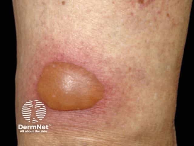
Fixed drug eruption can be categorised by clinical morphology. The most common form is the localised pigmenting type; other presentations include bullous (localised or generalised), mucosal, non-pigmenting, or generalised.

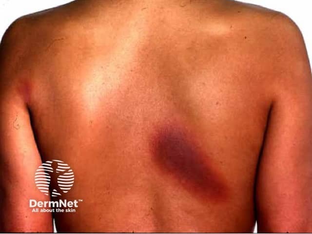
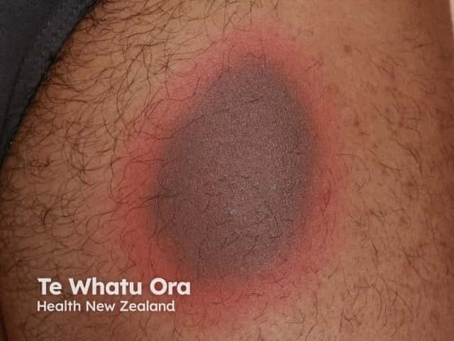
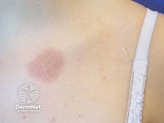
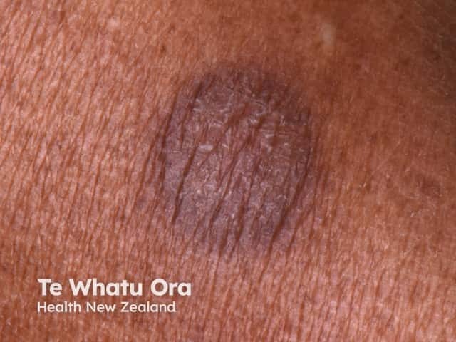
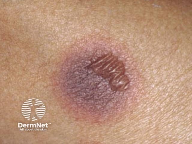
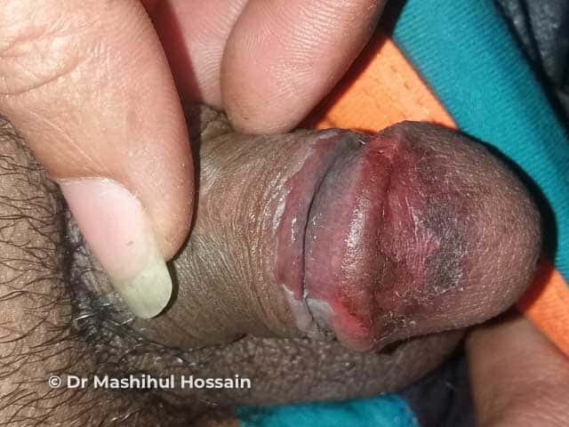
Fixed drug eruption typically presents as a single (or small number of) well-defined, round or oval red or violaceous patch or plaque which may blister or ulcerate. It is usually asymptomatic but can be itchy or painful. Over the next few days and weeks, the surface may become scaly or crusted before peeling, and the colour fades to leave brown post-inflammatory hyperpigmentation. Post-inflammatory hyperpigmentation tends to be more prominent in skin of colour.

In contrast to many other drug eruptions, the patient remains systemically well.

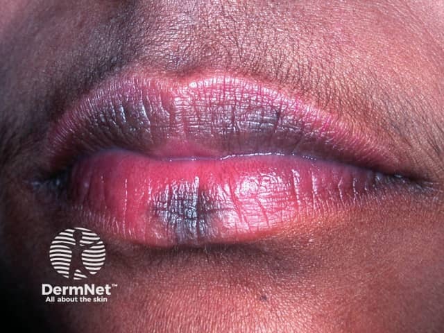
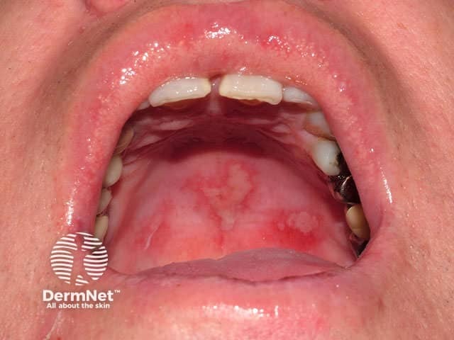
The hands and feet, eyelids, and anogenital areas are common sites. Lesions in the oral mucosa are usually found on the lips, tongue and hard palate. A fixed drug eruption may occur at the same location as previous skin trauma such as a burn, insect bite, or venepuncture.

On the first occasion, the eruption may develop after weeks to years of regular ingestion of the drug, but subsequent episodes develop within minutes to hours of recommencing the implicated drug. A patch of fixed drug eruption shows a refractory period during which it will not flare even with re-exposure. With subsequent episodes, the original patch may enlarge and more patches may appear. The post-inflammatory hyperpigmentation darkens with each recurrence.

Fixed drug eruption

Clinical variants of fixed drug eruption

Mucosal fixed drug eruption

  • Involves lips, tongue, hard palate, genital mucosa
  • Blisters and erosions are common
  • Can be isolated/localised or may occur with cutaneous lesions
  • Oral mucosal lesions commonly due to cotrimoxazole and naproxen
  • Genital mucosal lesions: glans penis – cotrimoxazole; vulvaNSAIDs.

Mucosal fixed drug eruption

Non-pigmenting fixed drug eruption

  • Often symmetrical lesions
  • Resolves without post-inflammatory hyperpigmentation
  • Associated with piroxicam and pseudoephedrine.

Generalised fixed drug eruption

  • Presents with numerous lesions
  • Lesions may be targetoid resembling erythema multiforme.

Generalised bullous fixed drug eruption

  • Rare variant
  • Recurrent episodes with onset within 24 hours of drug exposure
  • Numerous large blisters and erosions with normal skin between typically affecting <10% of the skin surface
  • Relative sparing of mucosal surfaces
  • Lesions are not targetoid
  • Fever, malaise and arthralgia may be associated
  • Resolves with post-inflammatory hyperpigmentation.

What are the complications of fixed drug eruption?

  • Blisters and erosions
  • Post-inflammatory hyperpigmentation
  • Recurrence
  • Cross-reaction with other medications
  • Generalised bullous fixed drug eruption can be complicated by fluid loss, electrolyte imbalance, and secondary infection.

How is fixed drug eruption diagnosed?

Fixed drug eruption should be considered on history and examination but may be difficult on the first occasion. On subsequent episodes, a detailed history of oral intake in the preceding 24 hours may identify the culprit.

Investigations may include:

  • Skin biopsy — shows an interface dermatitis in an early lesion with scattered apoptotic keratinocytes, vacuolar degeneration, dermal oedema, and a superficial perivascular lympho-eosinophilic infiltrate. Blisters are subepidermal if present. A late lesion shows upper dermal melanophages.
  • Oral challenge test — with a low dose of the suspected drug although there is typically a refractory period during which time the patch will not flare. This is contraindicated in patients with generalised bullous fixed drug eruption.
  • Patch test — using the suspected drug in soft paraffin applied to the lesion site is positive in 50% of cases. Prick testing and patch testing on normal skin is usually negative.

What is the differential diagnosis for fixed drug eruption?

What is the treatment for fixed drug eruption?

  • Discontinuation of suspected medication
  • Avoiding implicated medication indefinitely
  • Topical steroids/systemic corticosteroids
  • Generalised bullous fixed drug eruption requires intensive care or burns unit

What is the outcome for fixed drug eruption?

Fixed drug eruption is generally a benign self-resolving eruption that recurs on re-exposure, leaving post-inflammatory hyperpigmentation. Subsequent flares can be more severe.

Generalised bullous fixed drug eruption can be life-threatening, and has been reported to have a 20% mortality rate.

Appendix: list of some causes of fixed drug eruption

Antimicrobials

Analgesics/anti-inflammatories

Sedatives and anticonvulsants

Carbamazepine, barbiturates, and benzodiazepines

Antihypertensives

  • Calcium-channel blockers
  • Angiotensin-converting enzyme inhibitors

Other drugs

Cetirizine, omeprazole, pseudoephedrine, sulphasalazine, vaccinations

Complimentary medicines

Including traditional herbal medications and supplements

Foods

Asparagus, cashew nuts, kiwi fruit, lentil, palm wine, peanut, propolis, quinine (tonic water), seafood, strawberry, tartrazine (yellow rice, cheese crisps)


Bibliography

  • Adern-Jones MR, Lee HY. Benign cutaneous adverse reactions to drugs. In: Griffiths C, Barker J, Bleiker T, Chalmers R, Creamer D (eds). Rook’s Textbook of Dermatology, 9th edn. Wiley Blackwell, 2016: pp118.11–14.
  • Byrd RC, Mournighan KJ, Baca-Atlas M, Helton MR, Sun NZ, Siegel MB. Generalized bullous fixed-drug eruption secondary to the influenza vaccine. JAAD Case Rep. 2018;4(9):953–5. doi:10.1016/j.jdcr.2018.07.013. PubMed Central
  • Fukushima S, Kidou M, Ihn H. Fixed food eruption caused by cashew nut. Allergol Int. 2008;57(3):285–7. doi:10.2332/allergolint.C-07-58. Journal
  • Mitre V, Applebaum DS, Albahrani Y, Hsu S. Generalized bullous fixed drug eruption imitating toxic epidermal necrolysis: a case report and literature review. Dermatol Online J. 2017;23(7):13030/qt25v009gs. Journal
  • Mizukawa Y, Shiohara T. Trauma-localized fixed drug eruption: involvement of burn scars, insect bites and venipuncture sites. Dermatology. 2002;205(2):159–61. doi:10.1159/000063892. PubMed
  • Mizukawa Y, Yamazaki Y, Teraki Y, et al. Direct evidence for interferon-gamma production by effector-memory-type intraepidermal T cells residing at an effector site of immunopathology in fixed drug eruption. Am J Pathol. 2002;161(4):1337–47. doi:10.1016/s0002-9440(10)64410-0. Journal
  • Özkaya E. Oral mucosal fixed drug eruption: characteristics and differential diagnosis. J Am Acad Dermatol. 2013;69(2):e51–8. doi:10.1016/j.jaad.2012.08.019. PubMed
  • Patel S, John AM, Handler MZ, Schwartz RA. Fixed drug eruptions: an update, emphasizing the potentially lethal generalized bullous fixed drug eruption. Am J Clin Dermatol. 2020;21(3):393-9. doi:10.1007/s40257-020-00505-3. PubMed
  • Tan C, Zhu WY. Chinese herbal medicine: a neglected offender for fixed drug eruptions. Eur J Dermatol. 2010;20(3):397–9. doi:10.1684/ejd.2010.0947. PubMed

On DermNet

Other websites

Books about skin diseases


Related information

Sign up to the newsletter